2R5Q

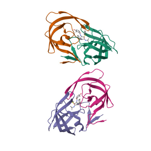
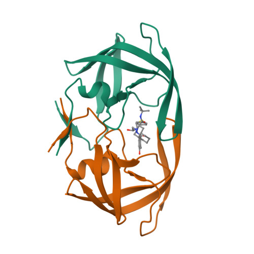
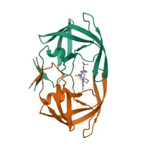
Crystal Structure Analysis of HIV-1 Subtype C Protease Complexed with Nelfinavir


Experimental Data Snapshot

  • Method: X-RAY DIFFRACTION
  • Resolution: 2.30 Å
  • R-Value Free: 0.283 
  • R-Value Work: 0.253 

wwPDB Validation   3D Report Full Report


Ligand Structure Quality Assessment 


This is version 1.4 of the entry. See complete history


Literature

The Contribution of Naturally Occurring Polymorphisms in Altering the Biochemical and Structural Characteristics of HIV-1 Subtype C Protease

Coman, R.M.Robbins, A.H.Fernandez, M.A.Gilliland, C.T.Sochet, A.A.Goodenow, M.M.McKenna, R.Dunn, B.M.

(2008) Biochemistry 47: 731-743

  • DOI: https://doi.org/10.1021/bi7018332
  • Primary Citation of Related Structures:  
    2R5P, 2R5Q

  • PubMed Abstract: 

    Fourteen subtype B and C protease variants have been engineered in an effort to study whether the preexistent baseline polymorphisms, by themselves or in combination with drug resistance mutations, differentially alter the biochemical and structural features of the subtype C protease when compared with those of subtype B protease. The kinetic studies performed in this work showed that the preexistent polymorphisms in subtype C protease, by themselves, do not provide for a greater level of resistance. Inhibition analysis with eight clinically used protease inhibitors revealed that the natural polymorphisms found in subtype C protease, in combination with drug resistance mutations, can influence enzymatic catalytic efficiency and inhibitor resistance. Structural analyses of the subtype C protease bound to nelfinavir and indinavir showed that these inhibitors form similar interactions with the residues in the active site of subtype B and C proteases. It also revealed that the naturally occurring polymorphisms could alter the position of the outer loops of the subtype C protease, especially the 60's loop.


  • Organizational Affiliation

    Department of Biochemistry and Molecular Biology, University of Florida College of Medicine, Gainesville, Florida 32610, USA


Macromolecules
Find similar proteins by:  (by identity cutoff)  |  3D Structure
Entity ID: 1
MoleculeChains Sequence LengthOrganismDetailsImage
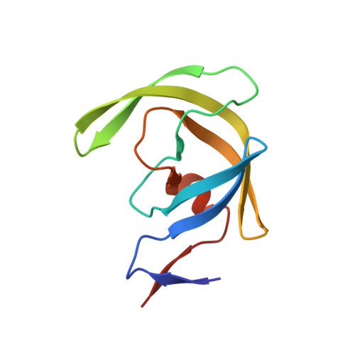
Protease
A, B, C, D
99Human immunodeficiency virus 1Mutation(s): 3 
Gene Names: pol
UniProt
Find proteins for O12158 (Human immunodeficiency virus type 1 group M subtype C (isolate 92BR025))
Explore O12158 
Go to UniProtKB:  O12158
Entity Groups  
Sequence Clusters30% Identity50% Identity70% Identity90% Identity95% Identity100% Identity
UniProt GroupO12158
Sequence Annotations
Expand
  • Reference Sequence
Small Molecules
Ligands 1 Unique
IDChains Name / Formula / InChI Key2D Diagram3D Interactions
1UN
Query on 1UN

Download Ideal Coordinates CCD File 
E [auth B],
F [auth D]
2-[2-HYDROXY-3-(3-HYDROXY-2-METHYL-BENZOYLAMINO)-4-PHENYL SULFANYL-BUTYL]-DECAHYDRO-ISOQUINOLINE-3-CARBOXYLIC ACID TERT-BUTYLAMIDE
C32 H45 N3 O4 S
QAGYKUNXZHXKMR-HKWSIXNMSA-N
Binding Affinity Annotations 
IDSourceBinding Affinity
NFV PDBBind:  2R5Q Ki: 2.7 (nM) from 1 assay(s)
Experimental Data & Validation

Experimental Data

  • Method: X-RAY DIFFRACTION
  • Resolution: 2.30 Å
  • R-Value Free: 0.283 
  • R-Value Work: 0.253 
  • Space Group: P 1 21 1
Unit Cell:
Length ( Å )Angle ( ˚ )
a = 46.717α = 90
b = 60.061β = 94.67
c = 86.71γ = 90
Software Package:
Software NamePurpose
DENZOdata reduction
SCALEPACKdata scaling
CNSrefinement
PDB_EXTRACTdata extraction
HKL-2000data collection
HKL-2000data reduction
HKL-2000data scaling
CNSphasing

Structure Validation

View Full Validation Report


Ligand Structure Quality Assessment 


Entry History 

Deposition Data

Revision History  (Full details and data files)

  • Version 1.0: 2007-11-20
    Type: Initial release
  • Version 1.1: 2011-07-13
    Changes: Version format compliance
  • Version 1.2: 2017-10-25
    Changes: Refinement description
  • Version 1.3: 2021-10-20
    Changes: Database references, Derived calculations
  • Version 1.4: 2024-02-21
    Changes: Data collection