2R1D | pdb_00002r1d

Crystal structure of rat neurexin 1beta in the Ca2+ containing form


Experimental Data Snapshot

  • Method: X-RAY DIFFRACTION
  • Resolution: 2.60 Å
  • R-Value Free: 
    0.244 (Depositor), 0.246 (DCC) 
  • R-Value Work: 
    0.203 (Depositor), 0.206 (DCC) 
  • R-Value Observed: 
    0.205 (Depositor) 

Starting Model: experimental
View more details

wwPDB Validation 3D Report Full Report

Validation slider image for 2R1D

This is version 1.3 of the entry. See complete history

Literature

Regulation of Neurexin 1beta Tertiary Structure and Ligand Binding through Alternative Splicing

Shen, K.C.Kuczynska, D.A.Wu, I.J.Murray, B.H.Sheckler, L.R.Rudenko, G.

(2008) Structure 16: 422-431

  • DOI: https://doi.org/10.1016/j.str.2008.01.005
  • Primary Citation Related Structures: 
    2R16, 2R1B, 2R1D

  • PubMed Abstract: 

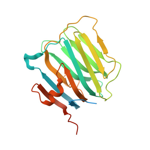
    Neurexins and neuroligins play an essential role in synapse function, and their alterations are linked to autistic spectrum disorder. Interactions between neurexins and neuroligins regulate inhibitory and excitatory synaptogenesis in vitro through a "splice-insert signaling code." In particular, neurexin 1beta carrying an alternative splice insert at site SS#4 interacts with neuroligin 2 (found predominantly at inhibitory synapses) but much less so with other neuroligins (those carrying an insert at site B and prevalent at excitatory synapses). The structure of neurexin 1beta+SS#4 reveals dramatic rearrangements to the "hypervariable surface," the binding site for neuroligins. The splice insert protrudes as a long helix into space, triggers conversion of loop beta10-beta11 into a helix rearranging the binding site for neuroligins, and rearranges the Ca(2+)-binding site required for ligand binding, increasing its affinity. Our structures reveal the mechanism by which neurexin 1beta isoforms acquire neuroligin splice isoform selectivity.


  • Organizational Affiliation
    • Life Sciences Institute, University of Michigan, Ann Arbor, MI 48109, USA.

Macromolecule Content 

  • Total Structure Weight: 243.33 kDa 
  • Atom Count: 12,217 
  • Modeled Residue Count: 1,600 
  • Deposited Residue Count: 2,260 
  • Unique protein chains: 1

Macromolecules

Find similar proteins by:|  3D Structure
Entity ID: 1
MoleculeChains  Sequence LengthOrganismDetailsImage
Neurexin-1-beta
A,
B,
C,
D,
E,
F,
G,
H,
I,
J [auth W]
226Rattus norvegicusMutation(s): 0 
Gene Names: Nrxn1
UniProt
Find proteins for Q63373 (Rattus norvegicus)
Explore Q63373 
Go to UniProtKB:  Q63373
Entity Groups
Sequence Clusters30% Identity50% Identity70% Identity90% Identity95% Identity100% Identity
UniProt GroupQ63373
Sequence Annotations
Expand
Reference Sequence

Experimental Data & Validation

Experimental Data

  • Method: X-RAY DIFFRACTION
  • Resolution: 2.60 Å
  • R-Value Free:  0.244 (Depositor), 0.246 (DCC) 
  • R-Value Work:  0.203 (Depositor), 0.206 (DCC) 
  • R-Value Observed: 0.205 (Depositor) 
Space Group: P 21 21 2
Unit Cell:
Length ( Å )Angle ( ˚ )
a = 116.683α = 90
b = 195.719β = 90
c = 103.867γ = 90
Software Package:
Software NamePurpose
REFMACrefinement
HKL-2000data reduction
HKL-2000data scaling

Structure Validation

View Full Validation Report


Entry History 

Deposition Data

  • Released Date: 2008-04-01 
  • Deposition Author(s): Rudenko, G.

Revision History  (Full details and data files)

  • Version 1.0: 2008-04-01
    Type: Initial release
  • Version 1.1: 2011-07-13
    Changes: Advisory, Version format compliance
  • Version 1.2: 2017-10-25
    Changes: Refinement description
  • Version 1.3: 2023-08-30
    Changes: Data collection, Database references, Derived calculations, Refinement description