


Crystal structure of putative Xaa-His dipeptidase (YP_718209.1) from Haemophilus somnus 129PT at 2.11 A resolution
Joint Center for Structural Genomics (JCSG)To be published.
Experimental Data Snapshot
wwPDB Validation 3D Report Full Report

Entity ID: 1 | |||||
|---|---|---|---|---|---|
| Molecule | Chains | Sequence Length | Organism | Details | Image |
| Xaa-His dipeptidase | 487 | Histophilus somni 129PT | Mutation(s): 0 Gene Names: pepD, HS_0004 EC: 3.4.13.20 (PDB Primary Data), 3.4.13.18 (UniProt) | 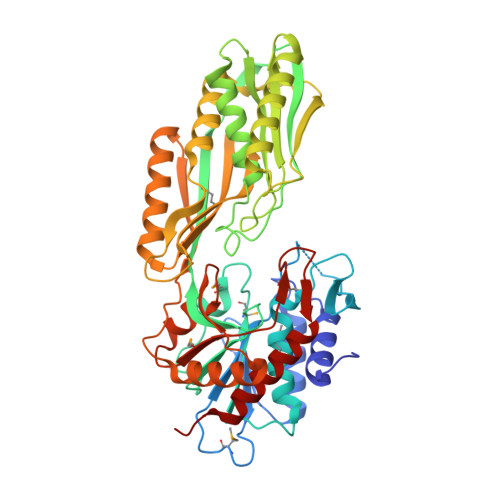 | |
UniProt | |||||
Entity Groups | |||||
| Sequence Clusters | 30% Identity50% Identity70% Identity90% Identity95% Identity100% Identity | ||||
| UniProt Group | Q0I1B9 | ||||
Sequence AnnotationsExpand | |||||
Reference Sequence | |||||
| Ligands 4 Unique | |||||
|---|---|---|---|---|---|
| ID | Chains | Name / Formula / InChI Key | 2D Diagram | 3D Interactions | |
| SO4 Download:Ideal Coordinates CCD File | AA [auth B] BA [auth B] CA [auth B] DA [auth B] E [auth A] | SULFATE ION O4 S QAOWNCQODCNURD-UHFFFAOYSA-L |  | ||
| GOL Download:Ideal Coordinates CCD File | IA [auth B] JA [auth B] KA [auth B] LA [auth B] M [auth A] | GLYCEROL C3 H8 O3 PEDCQBHIVMGVHV-UHFFFAOYSA-N |  | ||
| ZN Download:Ideal Coordinates CCD File | C [auth A], D [auth A], W [auth B], X [auth B] | ZINC ION Zn PTFCDOFLOPIGGS-UHFFFAOYSA-N |  | ||
| IPA Download:Ideal Coordinates CCD File | RA [auth B], U [auth A], V [auth A] | ISOPROPYL ALCOHOL C3 H8 O KFZMGEQAYNKOFK-UHFFFAOYSA-N |  | ||
| Modified Residues 1 Unique | |||||
|---|---|---|---|---|---|
| ID | Chains | Type | Formula | 2D Diagram | Parent |
| MSE Query on MSE | A, B | L-PEPTIDE LINKING | C5 H11 N O2 Se |  | MET |
| Length ( Å ) | Angle ( ˚ ) |
|---|---|
| a = 173.922 | α = 90 |
| b = 84.293 | β = 90 |
| c = 123.204 | γ = 90 |
| Software Name | Purpose |
|---|---|
| REFMAC | refinement |
| PHENIX | refinement |
| SHELX | phasing |
| MolProbity | model building |
| SCALA | data scaling |
| PDB_EXTRACT | data extraction |
| MAR345 | data collection |
| XDS | data reduction |
| autoSHARP | phasing |


