2QXV | pdb_00002qxv

Structural basis of EZH2 recognition by EED


Experimental Data Snapshot

  • Method: X-RAY DIFFRACTION
  • Resolution: 1.82 Å
  • R-Value Free: 
    0.218 (Depositor) 
  • R-Value Work: 
    0.185 (Depositor), 0.189 (DCC) 

wwPDB Validation 3D Report Full Report

Validation slider image for 2QXV

This is version 1.2 of the entry. See complete history

Literature

Structural basis of EZH2 recognition by EED

Han, Z.Xing, X.Hu, M.Zhang, Y.Liu, P.Chai, J.

(2007) Structure 15: 1306-1315

  • DOI: https://doi.org/10.1016/j.str.2007.08.007
  • Primary Citation Related Structures: 
    2QXV

  • PubMed Abstract: 

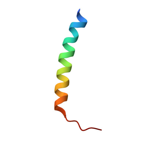
    The WD-repeat domain is a highly conserved recognition module in eukaryotes involved in diverse cellular processes. It is still not well understood how the bottom of a WD-repeat domain recognizes its binding partners. The WD-repeat-containing protein EED is one component of the PRC2 complex that possesses histone methyltransferase activity required for gene repression. Here we report the crystal structure of EED in complex with a 30 residue peptide from EZH2. The structure reveals that the peptide binds to the bottom of the WD-repeat domain of EED. The structural determinants of EZH2-EED interaction are present not only in EZH2 and EZH1 but also in its Drosophila homolog E(Z), suggesting that the recognition of ESC by E(Z) in Drosophila employs similar structural motifs. Structure-based mutagenesis identified critical residues from both EED and EZH2 for their interaction. The structure presented here may provide a template for understanding of how WD-repeat proteins recognize their interacting proteins.


  • Organizational Affiliation
    • National Institute of Biological Sciences, Beijing 102206, China.

Macromolecule Content 

  • Total Structure Weight: 45.4 kDa 
  • Atom Count: 3,493 
  • Modeled Residue Count: 381 
  • Deposited Residue Count: 391 
  • Unique protein chains: 2

Macromolecules

Find similar proteins by:|  3D Structure
Entity ID: 1
MoleculeChains  Sequence LengthOrganismDetailsImage
Embryonic ectoderm development361Mus musculusMutation(s): 0 
Gene Names: hEED
UniProt
Find proteins for Q921E6 (Mus musculus)
Explore Q921E6 
Go to UniProtKB:  Q921E6
Entity Groups
Sequence Clusters30% Identity50% Identity70% Identity90% Identity95% Identity100% Identity
UniProt GroupQ921E6
Sequence Annotations
Expand
Reference Sequence
Find similar proteins by:|  3D Structure
Entity ID: 2
MoleculeChains  Sequence LengthOrganismDetailsImage
Enhancer of zeste homolog 230Mus musculusMutation(s): 0 
Gene Names: Ezh2Enx1h
EC: 2.1.1.43 (PDB Primary Data), 2.1.1.356 (UniProt)
UniProt & NIH Common Fund Data Resources
Find proteins for Q61188 (Mus musculus)
Explore Q61188 
Go to UniProtKB:  Q61188
IMPC:  MGI:107940
Entity Groups
Sequence Clusters30% Identity50% Identity70% Identity90% Identity95% Identity100% Identity
UniProt GroupQ61188
Sequence Annotations
Expand
Reference Sequence

Experimental Data & Validation

Experimental Data

  • Method: X-RAY DIFFRACTION
  • Resolution: 1.82 Å
  • R-Value Free:  0.218 (Depositor) 
  • R-Value Work:  0.185 (Depositor), 0.189 (DCC) 
Space Group: P 21 21 21
Unit Cell:
Length ( Å )Angle ( ˚ )
a = 50.11α = 90
b = 52.74β = 90
c = 127.74γ = 90
Software Package:
Software NamePurpose
HKL-2000data collection
SOLVEphasing
CNSrefinement
DENZOdata reduction
SCALEPACKdata scaling

Structure Validation

View Full Validation Report


Entry History 

Deposition Data

  • Released Date: 2007-08-28 
  • Deposition Author(s): Han, Z.

Revision History  (Full details and data files)

  • Version 1.0: 2007-08-28
    Type: Initial release
  • Version 1.1: 2011-07-13
    Changes: Version format compliance
  • Version 1.2: 2024-03-13
    Changes: Data collection, Database references