2QUS | pdb_00002qus

Hammerhead Ribozyme G12A mutant pre-cleavage


Experimental Data Snapshot

  • Method: X-RAY DIFFRACTION
  • Resolution: 2.40 Å
  • R-Value Free: 
    0.253 (Depositor), 0.259 (DCC) 
  • R-Value Work: 
    0.184 (Depositor), 0.192 (DCC) 
  • R-Value Observed: 
    0.191 (Depositor) 

Starting Model: in silico
View more details

wwPDB Validation 3D Report Full Report

Validation slider image for 2QUS

This is version 1.3 of the entry. See complete history

Literature

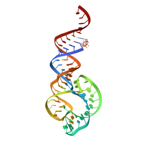
Capturing hammerhead ribozyme structures in action by modulating general base catalysis.

Chi, Y.I.Martick, M.Lares, M.Kim, R.Scott, W.G.Kim, S.H.

(2008) PLoS Biol 6: e234-e234

  • DOI: https://doi.org/10.1371/journal.pbio.0060234
  • Primary Citation Related Structures: 
    2QUS, 2QUW

  • PubMed Abstract: 

    We have obtained precatalytic (enzyme-substrate complex) and postcatalytic (enzyme-product complex) crystal structures of an active full-length hammerhead RNA that cleaves in the crystal. Using the natural satellite tobacco ringspot virus hammerhead RNA sequence, the self-cleavage reaction was modulated by substituting the general base of the ribozyme, G12, with A12, a purine variant with a much lower pKa that does not significantly perturb the ribozyme's atomic structure. The active, but slowly cleaving, ribozyme thus permitted isolation of enzyme-substrate and enzyme-product complexes without modifying the nucleophile or leaving group of the cleavage reaction, nor any other aspect of the substrate. The predissociation enzyme-product complex structure reveals RNA and metal ion interactions potentially relevant to transition-state stabilization that are absent in precatalytic structures.


  • Organizational Affiliation
    • Department of Molecular and Cellular Biochemistry, Center for Structural Biology, University of Kentucky, Lexington, Kentucky, United States of America. ychi@uky.edu

Macromolecule Content 

  • Total Structure Weight: 45.2 kDa 
  • Atom Count: 3,113 
  • Modeled Residue Count: 138 
  • Deposited Residue Count: 138 
  • Unique nucleic acid chains: 1

Macromolecules

Find similar nucleic acids by:  (by identity cutoff) 
Entity ID: 1
MoleculeChains LengthOrganismImage
Hammerhead ribozyme
A, B
69N/A
Sequence Annotations
Expand
Reference Sequence

Small Molecules

Ligands 1 Unique
IDChains Name / Formula / InChI Key2D Diagram3D Interactions
MG

Query on MG


Download:Ideal Coordinates CCD File
C [auth A]
D [auth A]
E [auth A]
F [auth A]
G [auth A]
C [auth A],
D [auth A],
E [auth A],
F [auth A],
G [auth A],
H [auth A],
I [auth A],
J [auth A],
K [auth B],
L [auth B],
M [auth B],
N [auth B],
O [auth B],
P [auth B],
Q [auth B],
R [auth B]
MAGNESIUM ION
Mg
JLVVSXFLKOJNIY-UHFFFAOYSA-N

Experimental Data & Validation

Experimental Data

  • Method: X-RAY DIFFRACTION
  • Resolution: 2.40 Å
  • R-Value Free:  0.253 (Depositor), 0.259 (DCC) 
  • R-Value Work:  0.184 (Depositor), 0.192 (DCC) 
  • R-Value Observed: 0.191 (Depositor) 
Space Group: P 1
Unit Cell:
Length ( Å )Angle ( ˚ )
a = 27.926α = 74.57
b = 53.03β = 81.37
c = 71.963γ = 75.61
Software Package:
Software NamePurpose
REFMACrefinement
PDB_EXTRACTdata extraction

Structure Validation

View Full Validation Report


Entry History 

Deposition Data

Revision History  (Full details and data files)

  • Version 1.0: 2008-07-15
    Type: Initial release
  • Version 1.1: 2011-07-13
    Changes: Advisory, Version format compliance
  • Version 1.2: 2024-02-21
    Changes: Data collection, Database references, Derived calculations
  • Version 1.3: 2024-04-03
    Changes: Refinement description