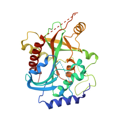
2QPL

Crystal structure of calf spleen purine nucleoside phosphorylase complexed to a novel purine analogue


Experimental Data Snapshot

  • Method: X-RAY DIFFRACTION
  • Resolution: 2.10 Å
  • R-Value Free: 0.218 
  • R-Value Work: 0.168 
  • R-Value Observed: 0.170 

Starting Model: experimental
View more details

wwPDB Validation   3D Report Full Report


Ligand Structure Quality Assessment 

Created with Raphaël 2.3.0Worse 01 BetterLigand structure goodness of fit to experimental dataBest fitted BTYClick on this verticalbar to view details

This is version 1.3 of the entry. See complete history


Literature

Crystal structure of calf spleen purine nucleoside phosphorylase complexed to a novel purine analogue.

Pereira, H.M.Berdini, V.Cleasby, A.Garratt, R.C.

(2007) FEBS Lett 581: 5082-5086

  • DOI: https://doi.org/10.1016/j.febslet.2007.09.051
  • Primary Citation of Related Structures:  
    2QPL

  • PubMed Abstract: 

    The combined use of a rapid virtual screen of a small fragment library together with a single point enzyme assay has been used for the discovery of novel PNP inhibitors. The availability of readily soakable crystals of bovine PNP has allowed the approach to be experimentally validated by determining the crystal structure of one of the inhibitor-PNP complexes. Comparison of the experimentally determined binding mode with that predicted by the virtual screening shows them to be similar. This represents a starting point for the growth of the ligand into a higher affinity inhibitor.


  • Organizational Affiliation

    Instituto de Física de São Carlos, Universidade de São Paulo, Physics and Informatics, Av. Trabalhador Saocarlense 400, Centro CP 369, 13560-970 Sao Carlos, São Paulo, Brazil. hmuniz.pereira@gmail.com


Macromolecules
Find similar proteins by:  (by identity cutoff)  |  3D Structure
Entity ID: 1
MoleculeChains Sequence LengthOrganismDetailsImage
Purine nucleoside phosphorylase282Bos taurusMutation(s): 0 
EC: 2.4.2.1
UniProt
Find proteins for P55859 (Bos taurus)
Explore P55859 
Go to UniProtKB:  P55859
Entity Groups  
Sequence Clusters30% Identity50% Identity70% Identity90% Identity95% Identity100% Identity
UniProt GroupP55859
Sequence Annotations
Expand
  • Reference Sequence
Experimental Data & Validation

Experimental Data

  • Method: X-RAY DIFFRACTION
  • Resolution: 2.10 Å
  • R-Value Free: 0.218 
  • R-Value Work: 0.168 
  • R-Value Observed: 0.170 
  • Space Group: P 21 3
Unit Cell:
Length ( Å )Angle ( ˚ )
a = 93.442α = 90
b = 93.442β = 90
c = 93.442γ = 90
Software Package:
Software NamePurpose
MOSFLMdata reduction
SCALAdata scaling
REFMACrefinement
PDB_EXTRACTdata extraction
ADSCdata collection
MOLREPphasing

Structure Validation

View Full Validation Report


Ligand Structure Quality Assessment 

Created with Raphaël 2.3.0Worse 01 BetterLigand structure goodness of fit to experimental dataBest fitted BTYClick on this verticalbar to view details

Entry History 

Deposition Data

Revision History  (Full details and data files)

  • Version 1.0: 2007-12-18
    Type: Initial release
  • Version 1.1: 2011-07-13
    Changes: Version format compliance
  • Version 1.2: 2023-08-30
    Changes: Data collection, Database references, Derived calculations, Refinement description
  • Version 1.3: 2023-11-29
    Changes: Database references