2QPK | pdb_00002qpk

Crystal structure of the complex of bovine lactoperoxidase with salicylhydroxamic acid at 2.34 A resolution


Experimental Data Snapshot

  • Method: X-RAY DIFFRACTION
  • Resolution: 2.34 Å
  • R-Value Free: 
    0.220 (Depositor), 0.220 (DCC) 
  • R-Value Work: 
    0.172 (Depositor), 0.176 (DCC) 
  • R-Value Observed: 
    0.174 (Depositor) 

Starting Model: experimental
View more details

wwPDB Validation 3D Report Full Report

Validation slider image for 2QPK

Ligand Structure Quality Assessment 


This is version 2.2 of the entry. See complete history

Literature

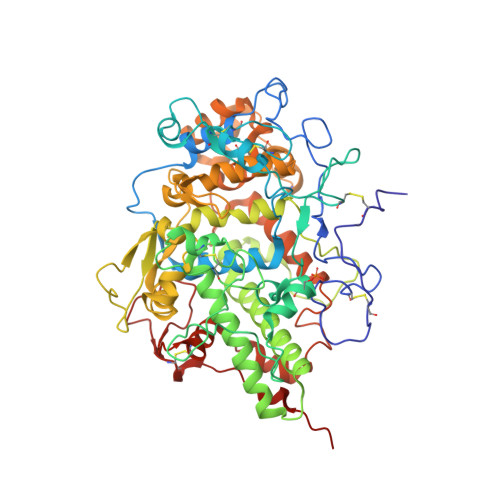
Structure of bovine lactoperoxidase with a partially linked heme moiety at 1.98 angstrom resolution.

Singh, P.K.Sirohi, H.V.Iqbal, N.Tiwari, P.Kaur, P.Sharma, S.Singh, T.P.

(2017) Biochim Biophys Acta 1865: 329-335

  • DOI: https://doi.org/10.1016/j.bbapap.2016.12.006
  • Primary Citation Related Structures: 
    2QPK, 3TGY, 4PNX, 5B72, 5GLS

  • PubMed Abstract: 

    Lactoperoxidase (LPO) is a member of mammalian heme peroxidase superfamily whose other members are myeloperoxidase (MPO), eosinophil peroxidase (EPO) and thyroid peroxidase (TPO). In these enzymes, the heme moiety is linked to protein through two or three covalent bonds. In the mature LPO, the heme moiety is linked to protein through two ester bonds with highly conserved glutamate and aspartate residues. The previously reported structures of LPO have confirmed the formation of two covalent linkages involving Glu258 and Asp108 with 1-methyl and 5-methyl groups of pyrrole rings A and C respectively. We report here a new form of structure of LPO where the covalent bond between Glu258 and 1-methyl group of pyrrole ring A is present only in a fraction of protein molecules. In this case, the side chain of Glu258 occupies two distinct positions, each of which has a 0.5 occupancy. In one position, it forms a normal ester covalent linkage while in the second position, the side chain of Glu258 is located in the middle of the substrate binding site on the distal heme side. In this position, the atom of the side chain of Glu258 forms several contacts with atoms of other residues and heme moiety. Out of the two observed positions of the side chain of Glu258, the former contributes to the stabilization of heme position and improved catalytic action of LPO while the latter is responsible for the reduced stability of the heme position as well as it blocks the substrate binding site.


  • Organizational Affiliation
    • Department of Biophysics, All India Institute of Medical Sciences, New Delhi, India.

Macromolecule Content 

  • Total Structure Weight: 71.76 kDa 
  • Atom Count: 5,275 
  • Modeled Residue Count: 595 
  • Deposited Residue Count: 595 
  • Unique protein chains: 1

Macromolecules

Find similar proteins by:|  3D Structure
Entity ID: 1
MoleculeChains  Sequence LengthOrganismDetailsImage
Lactoperoxidase595Bos taurusMutation(s): 0 
EC: 1.11.1.7
UniProt
Find proteins for P80025 (Bos taurus)
Explore P80025 
Go to UniProtKB:  P80025
Entity Groups
Sequence Clusters30% Identity50% Identity70% Identity90% Identity95% Identity100% Identity
UniProt GroupP80025
Glycosylation
Glycosylation Sites: 4
Sequence Annotations
Expand
Reference Sequence

Oligosaccharides

Help  
Entity ID: 2
MoleculeChains Length2D Diagram GlycosylationD Interactions
alpha-D-mannopyranose-(1-4)-2-acetamido-2-deoxy-beta-D-glucopyranose-(1-4)-2-acetamido-2-deoxy-beta-D-glucopyranose
B, D
3N-Glycosylation
Glycosylation Resources
GlyTouCan: G62182OO
GlyCosmos: G62182OO
GlyGen: G62182OO
Entity ID: 3
MoleculeChains Length2D Diagram GlycosylationD Interactions
2-acetamido-2-deoxy-beta-D-glucopyranose-(1-4)-2-acetamido-2-deoxy-beta-D-glucopyranose
C, E
2N-Glycosylation
Glycosylation Resources
GlyTouCan: G42666HT
GlyCosmos: G42666HT
GlyGen: G42666HT

Small Molecules

Ligands 5 Unique
IDChains Name / Formula / InChI Key2D Diagram3D Interactions
HEM

Query on HEM


Download:Ideal Coordinates CCD File
P [auth A]PROTOPORPHYRIN IX CONTAINING FE
C34 H32 Fe N4 O4
KABFMIBPWCXCRK-RGGAHWMASA-L
SHA

Query on SHA


Download:Ideal Coordinates CCD File
Q [auth A]SALICYLHYDROXAMIC ACID
C7 H7 N O3
HBROZNQEVUILML-UHFFFAOYSA-N
IOD

Query on IOD


Download:Ideal Coordinates CCD File
G [auth A]
H [auth A]
I [auth A]
J [auth A]
K [auth A]
G [auth A],
H [auth A],
I [auth A],
J [auth A],
K [auth A],
L [auth A],
M [auth A],
N [auth A]
IODIDE ION
I
XMBWDFGMSWQBCA-UHFFFAOYSA-M
SCN

Query on SCN


Download:Ideal Coordinates CCD File
O [auth A]THIOCYANATE ION
C N S
ZMZDMBWJUHKJPS-UHFFFAOYSA-M
CA

Query on CA


Download:Ideal Coordinates CCD File
F [auth A]CALCIUM ION
Ca
BHPQYMZQTOCNFJ-UHFFFAOYSA-N
Modified Residues  1 Unique
IDChains TypeFormula2D DiagramParent
SEP
Query on SEP
A
L-PEPTIDE LINKINGC3 H8 N O6 PSER
Binding Affinity Annotations 
IDSourceBinding Affinity
SHA BindingDB:  2QPK IC50: 400 (nM) from 1 assay(s)

Experimental Data & Validation

Experimental Data

  • Method: X-RAY DIFFRACTION
  • Resolution: 2.34 Å
  • R-Value Free:  0.220 (Depositor), 0.220 (DCC) 
  • R-Value Work:  0.172 (Depositor), 0.176 (DCC) 
  • R-Value Observed: 0.174 (Depositor) 
Space Group: P 1 21 1
Unit Cell:
Length ( Å )Angle ( ˚ )
a = 54.618α = 90
b = 80.553β = 102.56
c = 77.803γ = 90
Software Package:
Software NamePurpose
REFMACrefinement
MAR345dtbdata collection
DENZOdata reduction
SCALEPACKdata scaling
AMoREphasing

Structure Validation

View Full Validation Report


Ligand Structure Quality Assessment 


Entry History 

Deposition Data

Revision History  (Full details and data files)

  • Version 1.0: 2007-08-07
    Type: Initial release
  • Version 1.1: 2011-07-13
    Changes: Non-polymer description, Version format compliance
  • Version 1.2: 2017-02-22
    Changes: Database references
  • Version 2.0: 2020-07-29
    Type: Remediation
    Reason: Carbohydrate remediation
    Changes: Atomic model, Data collection, Derived calculations, Structure summary
  • Version 2.1: 2023-10-25
    Changes: Data collection, Database references, Refinement description, Structure summary
  • Version 2.2: 2024-10-09
    Changes: Structure summary