2QN1 | pdb_00002qn1

Glycogen Phosphorylase b in complex with asiatic acid


Experimental Data Snapshot

  • Method: X-RAY DIFFRACTION
  • Resolution: 2.40 Å
  • R-Value Free: 
    0.208 (Depositor), 0.207 (DCC) 
  • R-Value Work: 
    0.179 (Depositor), 0.178 (DCC) 
  • R-Value Observed: 
    0.181 (Depositor) 

Starting Model: experimental
View more details

wwPDB Validation 3D Report Full Report

Validation slider image for 2QN1

Ligand Structure Quality Assessment 


This is version 1.3 of the entry. See complete history

Literature

Naturally occurring pentacyclic triterpenes as inhibitors of glycogen phosphorylase: synthesis, structure-activity relationships, and X-ray crystallographic studies

Wen, X.Sun, H.Liu, J.Cheng, K.Zhang, P.Zhang, L.Hao, J.Zhang, L.Ni, P.Zographos, S.E.Leonidas, D.D.Alexacou, K.-M.Gimisis, T.Hayes, J.M.Oikonomakos, N.G.

(2008) J Med Chem 51: 3540-3554

  • DOI: https://doi.org/10.1021/jm8000949
  • Primary Citation Related Structures: 
    2QN1, 2QN2

  • PubMed Abstract: 

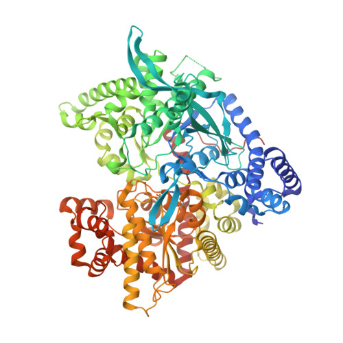
    Twenty-five naturally occurring pentacyclic triterpenes, 15 of which were synthesized in this study, were biologically evaluated as inhibitors of rabbit muscle glycogen phosphorylase a (GPa). From SAR studies, the presence of a sugar moiety in triterpene saponins resulted in a markedly decreased activity ( 7, 18- 20) or no activity ( 21, 22). These saponins, however, might find their value as potential natural prodrugs which are much more water-soluble than their corresponding aglycones. To elucidate the mechanism of GP inhibition, we have determined the crystal structures of the GPb-asiatic acid and GPb-maslinic acid complexes. The X-ray analysis indicates that the inhibitors bind at the allosteric activator site, where the physiological activator AMP binds. Pentacyclic triterpenes represent a promising class of multiple-target antidiabetic agents that exert hypoglycemic effects, at least in part, through GP inhibition.


  • Organizational Affiliation
    • Center for Drug Discovery, College of Pharmacy, China Pharmaceutical University, 24 Tongjia Xiang, Nanjing 210009, China.

Macromolecule Content 

  • Total Structure Weight: 98.01 kDa 
  • Atom Count: 6,871 
  • Modeled Residue Count: 813 
  • Deposited Residue Count: 842 
  • Unique protein chains: 1

Macromolecules

Find similar proteins by:|  3D Structure
Entity ID: 1
MoleculeChains  Sequence LengthOrganismDetailsImage
Glycogen phosphorylase, muscle form842Oryctolagus cuniculusMutation(s): 0 
EC: 2.4.1.1
UniProt
Find proteins for P00489 (Oryctolagus cuniculus)
Explore P00489 
Go to UniProtKB:  P00489
Entity Groups
Sequence Clusters30% Identity50% Identity70% Identity90% Identity95% Identity100% Identity
UniProt GroupP00489
Sequence Annotations
Expand
Reference Sequence

Small Molecules

Ligands 1 Unique
IDChains Name / Formula / InChI Key2D Diagram3D Interactions
0AS

Query on 0AS


Download:Ideal Coordinates CCD File
B [auth A]asiatic acid
C30 H48 O5
JXSVIVRDWWRQRT-UYDOISQJSA-N
Modified Residues  1 Unique
IDChains TypeFormula2D DiagramParent
LLP
Query on LLP
A
L-PEPTIDE LINKINGC14 H22 N3 O7 PLYS

Experimental Data & Validation

Experimental Data

  • Method: X-RAY DIFFRACTION
  • Resolution: 2.40 Å
  • R-Value Free:  0.208 (Depositor), 0.207 (DCC) 
  • R-Value Work:  0.179 (Depositor), 0.178 (DCC) 
  • R-Value Observed: 0.181 (Depositor) 
Space Group: P 43 21 2
Unit Cell:
Length ( Å )Angle ( ˚ )
a = 128.72α = 90
b = 128.72β = 90
c = 116.638γ = 90
Software Package:
Software NamePurpose
REFMACrefinement
DENZOdata reduction
SCALEPACKdata scaling

Structure Validation

View Full Validation Report


Ligand Structure Quality Assessment 


Entry History 

Deposition Data

Revision History  (Full details and data files)

  • Version 1.0: 2008-06-03
    Type: Initial release
  • Version 1.1: 2011-07-13
    Changes: Version format compliance
  • Version 1.2: 2023-08-30
    Changes: Data collection, Database references, Derived calculations, Refinement description
  • Version 1.3: 2023-11-15
    Changes: Data collection