2QB7 | pdb_00002qb7

Saccharomyces cerevisiae cytosolic exopolyphosphatase, phosphate complex


Experimental Data Snapshot

  • Method: X-RAY DIFFRACTION
  • Resolution: 1.60 Å
  • R-Value Free: 
    0.201 (Depositor) 
  • R-Value Work: 
    0.171 (Depositor) 
  • R-Value Observed: 
    0.172 (Depositor) 

Starting Model: experimental
View more details

wwPDB Validation 3D Report Full Report

Validation slider image for 2QB7

This is version 1.2 of the entry. See complete history

Literature

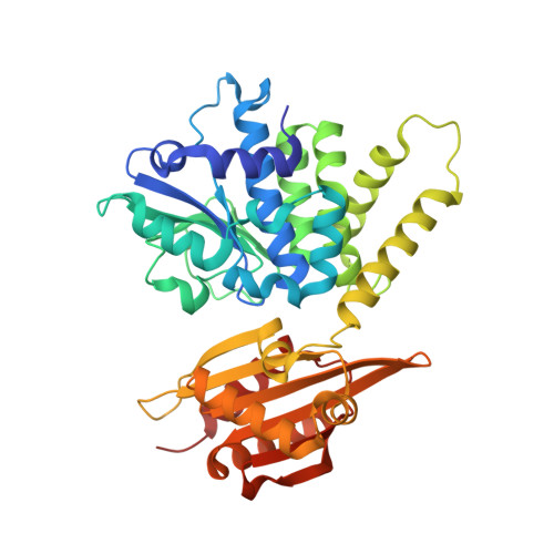
The crystal structure of the cytosolic exopolyphosphatase from Saccharomyces cerevisiae reveals the basis for substrate specificity.

Ugochukwu, E.Lovering, A.L.Mather, O.C.Young, T.W.White, S.A.

(2007) J Mol Biology 371: 1007-1021

  • DOI: https://doi.org/10.1016/j.jmb.2007.05.066
  • Primary Citation Related Structures: 
    2QB6, 2QB7, 2QB8

  • PubMed Abstract: 

    Inorganic long-chain polyphosphate is a ubiquitous linear polymer in biology, consisting of many phosphate moieties linked by phosphoanhydride bonds. It is synthesized by polyphosphate kinase, and metabolised by a number of enzymes, including exo- and endopolyphosphatases. The Saccharomyces cerevisiae gene PPX1 encodes for a 45 kDa, metal-dependent, cytosolic exopolyphosphatase that processively cleaves the terminal phosphate group from the polyphosphate chain, until inorganic pyrophosphate is all that remains. PPX1 belongs to the DHH family of phosphoesterases, which includes: family-2 inorganic pyrophosphatases, found in Gram-positive bacteria; prune, a cyclic AMPase; and RecJ, a single-stranded DNA exonuclease. We describe the high-resolution X-ray structures of yeast PPX1, solved using the multiple isomorphous replacement with anomalous scattering (MIRAS) technique, and its complexes with phosphate (1.6 A), sulphate (1.8 A) and ATP (1.9 A). Yeast PPX1 folds into two domains, and the structures reveal a strong similarity to the family-2 inorganic pyrophosphatases, particularly in the active-site region. A large, extended channel formed at the interface of the N and C-terminal domains is lined with positively charged amino acids and represents a conduit for polyphosphate and the site of phosphate hydrolysis. Structural comparisons with the inorganic pyrophosphatases and analysis of the ligand-bound complexes lead us to propose a hydrolysis mechanism. Finally, we discuss a structural basis for substrate selectivity and processivity.


  • Organizational Affiliation
    • The School of Biosciences, The University of Birmingham, Edgbaston, Birmingham, B15 2TT, UK.

Macromolecule Content 

  • Total Structure Weight: 91.45 kDa 
  • Atom Count: 7,133 
  • Modeled Residue Count: 782 
  • Deposited Residue Count: 794 
  • Unique protein chains: 1

Macromolecules

Find similar proteins by:|  3D Structure
Entity ID: 1
MoleculeChains  Sequence LengthOrganismDetailsImage
Exopolyphosphatase
A, B
397Saccharomyces cerevisiaeMutation(s): 0 
Gene Names: PPX1YHR201C
EC: 3.6.1.11
UniProt
Find proteins for P38698 (Saccharomyces cerevisiae (strain ATCC 204508 / S288c))
Explore P38698 
Go to UniProtKB:  P38698
Entity Groups
Sequence Clusters30% Identity50% Identity70% Identity90% Identity95% Identity100% Identity
UniProt GroupP38698
Sequence Annotations
Expand
Reference Sequence

Small Molecules

Ligands 4 Unique
IDChains Name / Formula / InChI Key2D Diagram3D Interactions
PO4

Query on PO4


Download:Ideal Coordinates CCD File
E [auth A]
F [auth A]
G [auth A]
M [auth B]
N [auth B]
E [auth A],
F [auth A],
G [auth A],
M [auth B],
N [auth B],
O [auth B]
PHOSPHATE ION
O4 P
NBIIXXVUZAFLBC-UHFFFAOYSA-K
EDO

Query on EDO


Download:Ideal Coordinates CCD File
J [auth A],
Q [auth B],
R [auth B],
S [auth B]
1,2-ETHANEDIOL
C2 H6 O2
LYCAIKOWRPUZTN-UHFFFAOYSA-N
ACT

Query on ACT


Download:Ideal Coordinates CCD File
H [auth A],
I [auth A],
P [auth B]
ACETATE ION
C2 H3 O2
QTBSBXVTEAMEQO-UHFFFAOYSA-M
CO

Query on CO


Download:Ideal Coordinates CCD File
C [auth A],
D [auth A],
K [auth B],
L [auth B]
COBALT (II) ION
Co
XLJKHNWPARRRJB-UHFFFAOYSA-N

Experimental Data & Validation

Experimental Data

  • Method: X-RAY DIFFRACTION
  • Resolution: 1.60 Å
  • R-Value Free:  0.201 (Depositor) 
  • R-Value Work:  0.171 (Depositor) 
  • R-Value Observed: 0.172 (Depositor) 
Space Group: P 21 21 21
Unit Cell:
Length ( Å )Angle ( ˚ )
a = 79.992α = 90
b = 83.039β = 90
c = 118.9γ = 90
Software Package:
Software NamePurpose
REFMACrefinement
ADSCdata collection
MOSFLMdata reduction
SCALAdata scaling
REFMACphasing

Structure Validation

View Full Validation Report


Entry History 

Deposition Data

Revision History  (Full details and data files)

  • Version 1.0: 2007-12-11
    Type: Initial release
  • Version 1.1: 2011-07-13
    Changes: Version format compliance
  • Version 1.2: 2023-08-30
    Changes: Data collection, Database references, Derived calculations, Refinement description