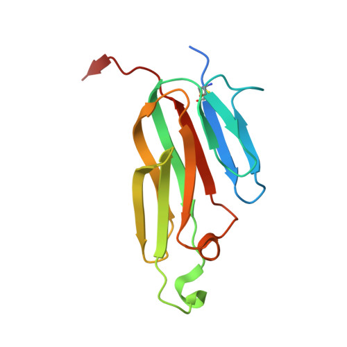
Structural basis for ligand-mediated mouse GITR activation.
Zhou, Z., Tone, Y., Song, X., Furuuchi, K., Lear, J.D., Waldmann, H., Tone, M., Greene, M.I., Murali, R.(2008) Proc Natl Acad Sci U S A 105: 641-645
- PubMed: 18178614
- DOI: https://doi.org/10.1073/pnas.0711206105
- Primary Citation of Related Structures:
2Q8O - PubMed Abstract:
Glucocorticoid-induced TNF receptor ligand (GITRL) is a member of the TNF super family (TNFSF). GITRL plays an important role in controlling regulatory T cells. The crystal structure of the mouse GITRL (mGITRL) was determined to 1.8-A resolution. Contrary to the current paradigm that all ligands in the TNFSF are trimeric, mGITRL associates as dimer through a unique C terminus tethering arm. Analytical ultracentrifuge studies revealed that in solution, the recombinant mGITRL exists as monomers at low concentrations and as dimers at high concentrations. Biochemical studies confirmed that the mGITRL dimer is biologically active. Removal of the three terminal residues in the C terminus resulted in enhanced receptor-mediated NF-kappaB activation than by the wild-type receptor complex. However, deletion of the tethering C-terminus arm led to reduced activity. Our studies suggest that the mGITRL may undergo a dynamic population shift among different oligomeric forms via C terminus-mediated conformational changes. We hypothesize that specific oligomeric forms of GITRL may be used as a means to differentially control GITR receptor signaling in diverse cells.
Organizational Affiliation:
Department of Pathology and Laboratory Medicine, University of Pennsylvania, Philadelphia, PA 19104, USA.


