2Q6G | pdb_00002q6g

Crystal structure of SARS-CoV main protease H41A mutant in complex with an N-terminal substrate


Experimental Data Snapshot

  • Method: X-RAY DIFFRACTION
  • Resolution: 2.50 Å
  • R-Value Free: 
    0.267 (Depositor), 0.266 (DCC) 
  • R-Value Work: 
    0.199 (Depositor), 0.200 (DCC) 

Starting Model: experimental
View more details

wwPDB Validation 3D Report Full Report

Validation slider image for 2Q6G

This is version 1.3 of the entry. See complete history

Literature

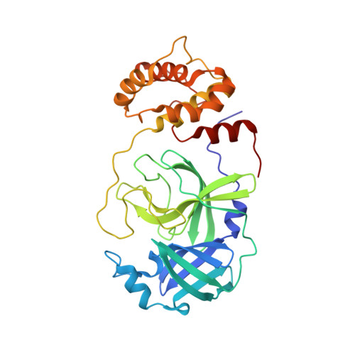
Structures of two coronavirus main proteases: implications for substrate binding and antiviral drug design.

Xue, X.Yu, H.Yang, H.Xue, F.Wu, Z.Shen, W.Li, J.Zhou, Z.Ding, Y.Zhao, Q.Zhang, X.C.Liao, M.Bartlam, M.Rao, Z.

(2008) J Virol 82: 2515-2527

  • DOI: https://doi.org/10.1128/JVI.02114-07
  • Primary Citation Related Structures: 
    2Q6D, 2Q6F, 2Q6G

  • PubMed Abstract: 

    Coronaviruses (CoVs) can infect humans and multiple species of animals, causing a wide spectrum of diseases. The coronavirus main protease (M(pro)), which plays a pivotal role in viral gene expression and replication through the proteolytic processing of replicase polyproteins, is an attractive target for anti-CoV drug design. In this study, the crystal structures of infectious bronchitis virus (IBV) M(pro) and a severe acute respiratory syndrome CoV (SARS-CoV) M(pro) mutant (H41A), in complex with an N-terminal autocleavage substrate, were individually determined to elucidate the structural flexibility and substrate binding of M(pro). A monomeric form of IBV M(pro) was identified for the first time in CoV M(pro) structures. A comparison of these two structures to other available M(pro) structures provides new insights for the design of substrate-based inhibitors targeting CoV M(pro)s. Furthermore, a Michael acceptor inhibitor (named N3) was cocrystallized with IBV M(pro) and was found to demonstrate in vitro inactivation of IBV M(pro) and potent antiviral activity against IBV in chicken embryos. This provides a feasible animal model for designing wide-spectrum inhibitors against CoV-associated diseases. The structure-based optimization of N3 has yielded two more efficacious lead compounds, N27 and H16, with potent inhibition against SARS-CoV M(pro).


  • Organizational Affiliation
    • Laboratory of Structural Biology, Life Sciences Building, Tsinghua University, Beijing 100084, China.

Macromolecule Content 

  • Total Structure Weight: 70.01 kDa 
  • Atom Count: 5,059 
  • Modeled Residue Count: 626 
  • Deposited Residue Count: 634 
  • Unique protein chains: 2

Macromolecules

Find similar proteins by:|  3D Structure
Entity ID: 1
MoleculeChains  Sequence LengthOrganismDetailsImage
severe acute respiratory syndrome coronavirus (SARS-CoV)
A, B
306SARS coronavirus BJ01Mutation(s): 1 
Gene Names: rep
EC: 3.4.22
UniProt
Find proteins for P0C6X7 (Severe acute respiratory syndrome coronavirus)
Explore P0C6X7 
Go to UniProtKB:  P0C6X7
Entity Groups
Sequence Clusters30% Identity50% Identity70% Identity90% Identity95% Identity100% Identity
UniProt GroupP0C6X7
Sequence Annotations
Expand
Reference Sequence
Find similar proteins by:  Sequence   |   3D Structure  
Entity ID: 2
MoleculeChains  Sequence LengthOrganismDetailsImage
Polypeptide chain
C, D
11N/AMutation(s): 0 
UniProt
Find proteins for P0C6X7 (Severe acute respiratory syndrome coronavirus)
Explore P0C6X7 
Go to UniProtKB:  P0C6X7
Entity Groups
Sequence Clusters30% Identity50% Identity70% Identity90% Identity95% Identity100% Identity
UniProt GroupP0C6X7
Sequence Annotations
Expand
Reference Sequence

Experimental Data & Validation

Experimental Data

  • Method: X-RAY DIFFRACTION
  • Resolution: 2.50 Å
  • R-Value Free:  0.267 (Depositor), 0.266 (DCC) 
  • R-Value Work:  0.199 (Depositor), 0.200 (DCC) 
Space Group: P 1 21 1
Unit Cell:
Length ( Å )Angle ( ˚ )
a = 51.982α = 90
b = 95.829β = 102.92
c = 67.718γ = 90
Software Package:
Software NamePurpose
HKL-2000data collection
CNSrefinement
HKL-2000data reduction
HKL-2000data scaling
CNSphasing

Structure Validation

View Full Validation Report


Entry History 

Deposition Data

Revision History  (Full details and data files)

  • Version 1.0: 2008-02-12
    Type: Initial release
  • Version 1.1: 2011-07-13
    Changes: Source and taxonomy, Version format compliance
  • Version 1.2: 2021-10-20
    Changes: Database references
  • Version 1.3: 2023-08-30
    Changes: Data collection, Refinement description