2Q55 | pdb_00002q55

Crystal structure of KK44 bound to HIV-1 protease


Experimental Data Snapshot

  • Method: X-RAY DIFFRACTION
  • Resolution: 1.90 Å
  • R-Value Free: 
    0.235 (Depositor), 0.244 (DCC) 
  • R-Value Work: 
    0.175 (Depositor), 0.198 (DCC) 
  • R-Value Observed: 
    0.178 (Depositor) 

Starting Model: experimental
View more details

wwPDB Validation 3D Report Full Report

Validation slider image for 2Q55

Ligand Structure Quality Assessment 


This is version 1.5 of the entry. See complete history

Literature

Design and Synthesis of HIV-1 Protease Inhibitors Incorporating Oxazolidinones as P2/P2' Ligands in Pseudosymmetric Dipeptide Isosteres.

Reddy, G.S.Ali, A.Nalam, M.N.Anjum, S.G.Cao, H.Nathans, R.S.Schiffer, C.A.Rana, T.M.

(2007) J Med Chem 50: 4316-4328

  • DOI: https://doi.org/10.1021/jm070284z
  • Primary Citation Related Structures: 
    2Q54, 2Q55, 2Q5K

  • PubMed Abstract: 

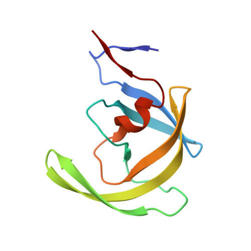
    A series of novel HIV-1 protease inhibitors based on two pseudosymmetric dipeptide isosteres have been synthesized and evaluated. The inhibitors were designed by incorporating N-phenyloxazolidinone-5-carboxamides into the hydroxyethylene and (hydroxyethyl)hydrazine dipeptide isosteres as P2 and P2' ligands. Compounds with (S)-phenyloxazolidinones attached at a position proximal to the central hydroxyl group showed low nM inhibitory activities against wild-type HIV-1 protease. Selected compounds were further evaluated for their inhibitory activities against a panel of multidrug-resistant protease variants and for their antiviral potencies in MT-4 cells. The crystal structures of lopinavir (LPV) and two new inhibitors containing phenyloxazolidinone-based ligands in complex with wild-type HIV-1 protease have been determined. A comparison of the inhibitor-protease structures with the LPV-protease structure provides valuable insight into the binding mode of the new inhibitors to the protease enzyme. Based on the crystal structures and knowledge of structure-activity relationships, new inhibitors can be designed with enhanced enzyme inhibitory and antiviral potencies.


  • Organizational Affiliation
    • Chemical Biology Program and Department of Biochemistry and Molecular Pharmacology, University of Massachusetts Medical School, Worcester, Massachusetts 01605, USA.

Macromolecule Content 

  • Total Structure Weight: 22.48 kDa 
  • Atom Count: 1,700 
  • Modeled Residue Count: 198 
  • Deposited Residue Count: 198 
  • Unique protein chains: 1

Macromolecules

Find similar proteins by:|  3D Structure
Entity ID: 1
MoleculeChains  Sequence LengthOrganismDetailsImage
Protease
A, B
99Human immunodeficiency virus 1Mutation(s): 1 
Gene Names: pol
UniProt
Find proteins for O38719 (Human immunodeficiency virus type 1)
Explore O38719 
Go to UniProtKB:  O38719
Entity Groups
Sequence Clusters30% Identity50% Identity70% Identity90% Identity95% Identity100% Identity
UniProt GroupO38719
Sequence Annotations
Expand
Reference Sequence

Small Molecules

Ligands 2 Unique
IDChains Name / Formula / InChI Key2D Diagram3D Interactions
MU0

Query on MU0


Download:Ideal Coordinates CCD File
E [auth B](5S)-N-[(1S,2S,4S)-1-BENZYL-2-HYDROXY-4-{[(2S)-3-METHYL-2-(2-OXOTETRAHYDROPYRIMIDIN-1(2H)-YL)BUTANOYL]AMINO}-5-PHENYLPENTYL]-2-OXO-3-PHENYL-1,3-OXAZOLIDINE-5-CARBOXAMIDE
C37 H45 N5 O6
YTEURAFEAFHROU-FLIXOAOSSA-N
PO4

Query on PO4


Download:Ideal Coordinates CCD File
C [auth A],
D [auth A]
PHOSPHATE ION
O4 P
NBIIXXVUZAFLBC-UHFFFAOYSA-K

Experimental Data & Validation

Experimental Data

  • Method: X-RAY DIFFRACTION
  • Resolution: 1.90 Å
  • R-Value Free:  0.235 (Depositor), 0.244 (DCC) 
  • R-Value Work:  0.175 (Depositor), 0.198 (DCC) 
  • R-Value Observed: 0.178 (Depositor) 
Space Group: P 21 21 21
Unit Cell:
Length ( Å )Angle ( ˚ )
a = 50.844α = 90
b = 58.53β = 90
c = 61.869γ = 90
Software Package:
Software NamePurpose
REFMACrefinement
HKL-2000data reduction
HKL-2000data scaling
AMoREphasing

Structure Validation

View Full Validation Report


Ligand Structure Quality Assessment 


Entry History 

Deposition Data

Revision History  (Full details and data files)

  • Version 1.0: 2007-09-04
    Type: Initial release
  • Version 1.1: 2011-07-13
    Changes: Derived calculations, Version format compliance
  • Version 1.2: 2017-10-18
    Changes: Refinement description
  • Version 1.3: 2021-10-20
    Changes: Database references, Derived calculations
  • Version 1.4: 2023-08-30
    Changes: Data collection, Refinement description
  • Version 1.5: 2024-03-13
    Changes: Source and taxonomy, Structure summary