2Q3Y | pdb_00002q3y

Ancestral Corticiod Receptor in Complex with DOC


Experimental Data Snapshot

  • Method: X-RAY DIFFRACTION
  • Resolution: 2.40 Å
  • R-Value Free: 
    0.240 (Depositor) 
  • R-Value Work: 
    0.196 (Depositor), 0.199 (DCC) 
  • R-Value Observed: 
    0.196 (Depositor) 

Starting Model: experimental
View more details

wwPDB Validation 3D Report Full Report

Validation slider image for 2Q3Y

Ligand Structure Quality Assessment 


This is version 1.3 of the entry. See complete history

Literature

Crystal structure of an ancient protein: evolution by conformational epistasis

Ortlund, E.A.Bridgham, J.T.Redinbo, M.R.Thornton, J.W.

(2007) Science 317: 1544-1548

  • DOI: https://doi.org/10.1126/science.1142819
  • Primary Citation Related Structures: 
    2Q1H, 2Q1V, 2Q3Y

  • PubMed Abstract: 

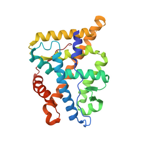
    The structural mechanisms by which proteins have evolved new functions are known only indirectly. We report x-ray crystal structures of a resurrected ancestral protein-the approximately 450 million-year-old precursor of vertebrate glucocorticoid (GR) and mineralocorticoid (MR) receptors. Using structural, phylogenetic, and functional analysis, we identify the specific set of historical mutations that recapitulate the evolution of GR's hormone specificity from an MR-like ancestor. These substitutions repositioned crucial residues to create new receptor-ligand and intraprotein contacts. Strong epistatic interactions occur because one substitution changes the conformational position of another site. "Permissive" mutations-substitutions of no immediate consequence, which stabilize specific elements of the protein and allow it to tolerate subsequent function-switching changes-played a major role in determining GR's evolutionary trajectory.


  • Organizational Affiliation
    • Department of Chemistry, University of North Carolina, Chapel Hill, NC 27599, USA.

Macromolecule Content 

  • Total Structure Weight: 30.41 kDa 
  • Atom Count: 2,196 
  • Modeled Residue Count: 254 
  • Deposited Residue Count: 259 
  • Unique protein chains: 2

Macromolecules

Find similar proteins by:|  3D Structure
Entity ID: 1
MoleculeChains  Sequence LengthOrganismDetailsImage
Ancestral Corticiod Receptor249unidentifiedMutation(s): 0 
Find similar proteins by:  Sequence   |   3D Structure  
Entity ID: 2
MoleculeChains  Sequence LengthOrganismDetailsImage
Nuclear receptor 0B210Homo sapiensMutation(s): 0 
Gene Names: NR0B2
UniProt & NIH Common Fund Data Resources
Find proteins for Q15466 (Homo sapiens)
Explore Q15466 
Go to UniProtKB:  Q15466
PHAROS:  Q15466
GTEx:  ENSG00000131910 
Entity Groups
Sequence Clusters30% Identity50% Identity70% Identity90% Identity95% Identity100% Identity
UniProt GroupQ15466
Sequence Annotations
Expand
Reference Sequence

Experimental Data & Validation

Experimental Data

  • Method: X-RAY DIFFRACTION
  • Resolution: 2.40 Å
  • R-Value Free:  0.240 (Depositor) 
  • R-Value Work:  0.196 (Depositor), 0.199 (DCC) 
  • R-Value Observed: 0.196 (Depositor) 
Space Group: P 43 21 2
Unit Cell:
Length ( Å )Angle ( ˚ )
a = 78.995α = 90
b = 78.995β = 90
c = 112.467γ = 90
Software Package:
Software NamePurpose
CNSrefinement
Blu-Icedata collection
HKL-2000data reduction
HKL-2000data scaling
PHASERphasing

Structure Validation

View Full Validation Report


Ligand Structure Quality Assessment 


Entry History 

Deposition Data

Revision History  (Full details and data files)

  • Version 1.0: 2007-09-04
    Type: Initial release
  • Version 1.1: 2011-07-13
    Changes: Non-polymer description, Version format compliance
  • Version 1.2: 2024-02-21
    Changes: Data collection, Database references, Derived calculations
  • Version 1.3: 2024-04-03
    Changes: Refinement description