2Q3N | pdb_00002q3n

Agglutinin from Abrus Precatorius (APA-I)


Experimental Data Snapshot

  • Method: X-RAY DIFFRACTION
  • Resolution: 3.50 Å
  • R-Value Free: 
    0.255 (Depositor), 0.213 (DCC) 
  • R-Value Work: 
    0.195 (Depositor) 
  • R-Value Observed: 
    0.201 (Depositor) 

Starting Model: experimental
View more details

wwPDB Validation 3D Report Full Report

Validation slider image for 2Q3N

Ligand Structure Quality Assessment 


This is version 1.5 of the entry. See complete history

Literature

Structure-Function Analysis and Insights into the Reduced Toxicity of Abrus precatorius Agglutinin I in Relation to Abrin.

Bagaria, A.Surendranath, K.Ramagopal, U.A.Ramakumar, S.Karande, A.A.

(2006) J Biological Chem 281: 34465-34474

  • DOI: https://doi.org/10.1074/jbc.M601777200
  • Primary Citation Related Structures: 
    2Q3N

  • PubMed Abstract: 

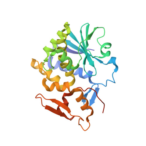
    Abrin and agglutinin-I from the seeds of Abrus precatorius are type II ribosome-inactivating proteins that inhibit protein synthesis in eukaryotic cells. The two toxins share a high degree of sequence similarity; however, agglutinin-I is weaker in its activity. We compared the kinetics of protein synthesis inhibition by abrin and agglutinin-I in two different cell lines and found that approximately 200-2000-fold higher concentration of agglutinin-I is needed for the same degree of inhibition. Like abrin, agglutinin-I also induced apoptosis in the cells by triggering the intrinsic mitochondrial pathway, although at higher concentrations as compared with abrin. The reason for the decreased toxicity of agglutinin-I became apparent on the analysis of the crystal structure of agglutinin-I obtained by us in comparison with that of the reported structure of abrin. The overall protein folding of agglutinin-I is similar to that of abrin-a with a single disulfide bond holding the toxic A subunit and the lectin-like B-subunit together, constituting a heterodimer. However, there are significant differences in the secondary structural elements, mostly in the A chain. The substitution of Asn-200 in abrin-a with Pro-199 in agglutinin-I seems to be a major cause for the decreased toxicity of agglutinin-I. This perhaps is not a consequence of any kink formation by a proline residue in the helical segment, as reported by others earlier, but due to fewer interactions that proline can possibly have with the bound substrate.


  • Organizational Affiliation
    • Department of Biochemistry, Indian Institute of Science, Bangalore 560012, India.

Macromolecule Content 

  • Total Structure Weight: 59.58 kDa 
  • Atom Count: 4,028 
  • Modeled Residue Count: 508 
  • Deposited Residue Count: 527 
  • Unique protein chains: 2

Macromolecules

Find similar proteins by:|  3D Structure
Entity ID: 1
MoleculeChains  Sequence LengthOrganismDetailsImage
Agglutinin-1 A chain260Abrus precatoriusMutation(s): 0 
EC: 3.2.2.22
UniProt
Find proteins for Q9M6E9 (Abrus precatorius)
Explore Q9M6E9 
Go to UniProtKB:  Q9M6E9
Entity Groups
Sequence Clusters30% Identity50% Identity70% Identity90% Identity95% Identity100% Identity
UniProt GroupQ9M6E9
Sequence Annotations
Expand
Reference Sequence
Find similar proteins by:|  3D Structure
Entity ID: 2
MoleculeChains  Sequence LengthOrganismDetailsImage
Agglutinin-1 B chain267Abrus precatoriusMutation(s): 0 
UniProt
Find proteins for Q9M6E9 (Abrus precatorius)
Explore Q9M6E9 
Go to UniProtKB:  Q9M6E9
Entity Groups
Sequence Clusters30% Identity50% Identity70% Identity90% Identity95% Identity100% Identity
UniProt GroupQ9M6E9
Sequence Annotations
Expand
Reference Sequence

Experimental Data & Validation

Experimental Data

  • Method: X-RAY DIFFRACTION
  • Resolution: 3.50 Å
  • R-Value Free:  0.255 (Depositor), 0.213 (DCC) 
  • R-Value Work:  0.195 (Depositor) 
  • R-Value Observed: 0.201 (Depositor) 
Space Group: P 42 21 2
Unit Cell:
Length ( Å )Angle ( ˚ )
a = 141.122α = 90
b = 141.122β = 90
c = 105.253γ = 90
Software Package:
Software NamePurpose
REFMACrefinement
HKL-2000data collection
HKL-2000data reduction
SCALEPACKdata scaling
AMoREphasing

Structure Validation

View Full Validation Report


Ligand Structure Quality Assessment 


Entry History 

Deposition Data

Revision History  (Full details and data files)

  • Version 1.0: 2007-06-26
    Type: Initial release
  • Version 1.1: 2008-05-01
    Changes: Version format compliance
  • Version 1.2: 2011-07-13
    Changes: Advisory, Derived calculations, Version format compliance
  • Version 1.3: 2020-07-29
    Type: Remediation
    Reason: Carbohydrate remediation
    Changes: Data collection, Derived calculations, Structure summary
  • Version 1.4: 2023-08-30
    Changes: Data collection, Database references, Refinement description, Structure summary
  • Version 1.5: 2024-10-30
    Changes: Structure summary