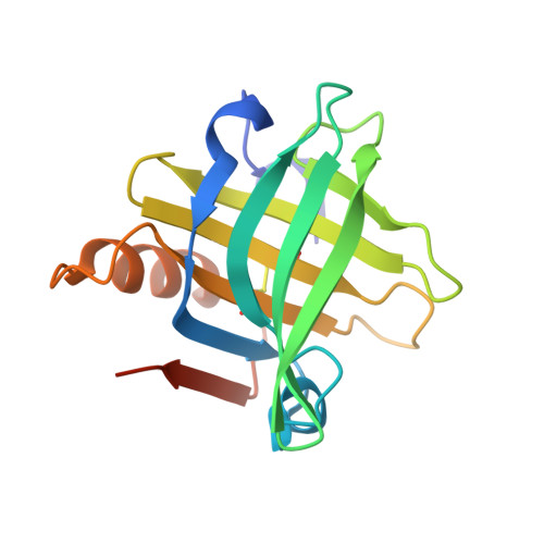
An asymmetric dimer of beta-lactoglobulin in a low humidity crystal form-Structural changes that accompany partial dehydration and protein action.
Vijayalakshmi, L., Krishna, R., Sankaranarayanan, R., Vijayan, M.(2007) Proteins 71: 241-249
- PubMed: 17932936
- DOI: https://doi.org/10.1002/prot.21695
- Primary Citation of Related Structures:
2Q2M, 2Q2P, 2Q39 - PubMed Abstract:
Dimeric lactoglobulin molecules exist in the open conformation at basic pH, whereas they exist in the closed conformation at acidic pH, after undergoing the Tanford transition around neutral pH. Orthorhombic crystals consisting of molecules in the open conformation, grown close to neutral pH, undergo a water-mediated transformation when the relative humidity around the crystals is reduced. The two subunits in the dimer are related by a crystallographic twofold axis in the native crystals while the dimer is asymmetric in the low humidity form. Interestingly, one of the subunits in the dimer in the low humidity form is in an open conformation while the other is in a closed conformation. This is the first observation of such an asymmetric dimer. A hydrogen bond between the side chains of Gln35 and Tyr42 exists and the side chain of Glu89 is substantially buried in the closed subunit of the asymmetric unit, as in other structures with molecules in the closed conformation. However, the closure of the EF loop is not complete; its conformation can be described as half-closed. A comparison of different crystal structures of beta-lactoglobulin indicates that the conformation of the loops in the molecule is substantially influenced by other factors such as crystal packing, the pH, and the composition of the medium, while the change in the conformation of the EF loop follows the Tanford transition. The mutual disposition of the two subunits in the low humidity form is halfway between those in the open and closed structures. The present work further demonstrates that structural changes that occur during partial dehydration could mimic those that occur during the action of proteins.
Organizational Affiliation:
Molecular Biophysics Unit, Indian Institute of Science, Bangalore, India.


