2PYS | pdb_00002pys

Crystal Structure of a Five Site Mutated Cyanovirin-N with a Mannose Dimer Bound at 1.8 A Resolution


Experimental Data Snapshot

  • Method: X-RAY DIFFRACTION
  • Resolution: 1.80 Å
  • R-Value Free: 
    0.215 (Depositor), 0.183 (DCC) 
  • R-Value Work: 
    0.169 (Depositor), 0.191 (DCC) 
  • R-Value Observed: 
    0.171 (Depositor) 

Starting Model: experimental
View more details

wwPDB Validation 3D Report Full Report

Validation slider image for 2PYS

This is version 2.3 of the entry. See complete history. 

Literature

A Monovalent Mutant of Cyanovirin-N Provides Insight into the Role of Multiple Interactions with gp120 for Antiviral Activity.

Fromme, R., Katiliene, Z., Giomarelli, B., Bogani, F., Mahon, J.M., Mori, T., Fromme, P., Ghirlanda, G.

(2007) Biochemistry 46: 9199-9207

  • DOI: https://doi.org/10.1021/bi700666m
  • Primary Citation Related Structures: 
    2PYS, 2Z21

  • PubMed Abstract: 

    Cyanovirin-N (CV-N) is a 101 amino acid cyanobacterial lectin with potent antiviral activity against HIV, mediated by high-affinity binding to branched N-linked oligomannosides on the viral surface envelope protein gp120. The protein contains two carbohydrate-binding domains, A and B, each of which binds short oligomannosides independently in vitro. The interaction to gp120 could involve either a single domain or both domains simultaneously; it is not clear which mode would elicit the antiviral activity. The model is complicated by the formation of a domain-swapped dimer form, in which part of each domain is exchanged between two monomers, which contains four functional carbohydrate-binding domains. To clarify whether multivalent interactions with gp120 are necessary for the antiviral activity, we engineered a novel mutant, P51G-m4-CVN, in which the binding site on domain A has been knocked out; in addition, a [P51G] mutation prevents the formation of domain-swapped dimers under physiological conditions. Here, we present the crystal structures at 1.8 A of the free and of the dimannose-bound forms of P51G-m4-CVN, revealing a monomeric structure in which only domain B is bound to dimannose. P51G-m4-CVN binds gp120 with an affinity almost 2 orders of magnitude lower than wt CV-N and is completely inactive against HIV. The tight binding to gp120 is recovered in the domain-swapped version of P51G-m4-CVN, prepared under extreme conditions. Our findings show that the presence of at least two oligomannoside-binding sites, either by the presence of intact domains A and B or by formation of domain-swapped dimers, is essential for activity.


  • Organizational Affiliation: 
    • Department of Chemistry and Biochemistry, Arizona State University, Tempe, Arizona 85287-1604, USA.

Macromolecule Content 

  • Total Structure Weight: 24.58 kDa 
  • Atom Count: 1,769 
  • Modeled Residue Count: 204 
  • Deposited Residue Count: 218 
  • Unique protein chains: 1

Macromolecules

Find similar proteins by:|  3D Structure
Entity ID: 1
MoleculeChains  Sequence LengthOrganismDetailsImage
Cyanovirin-N
A, B
109Nostoc ellipsosporumMutation(s): 5 
Gene Names: CYANOVIRN-N
UniProt
Find proteins for P81180 (Nostoc ellipsosporum)
Explore P81180 
Go to UniProtKB:  P81180
Entity Groups
Sequence Clusters30% Identity50% Identity70% Identity90% Identity95% Identity100% Identity
UniProt GroupP81180
Sequence Annotations
Expand
Reference Sequence

Oligosaccharides

Help  
Entity ID: 2
MoleculeChains Length2D Diagram GlycosylationD Interactions
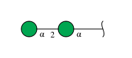
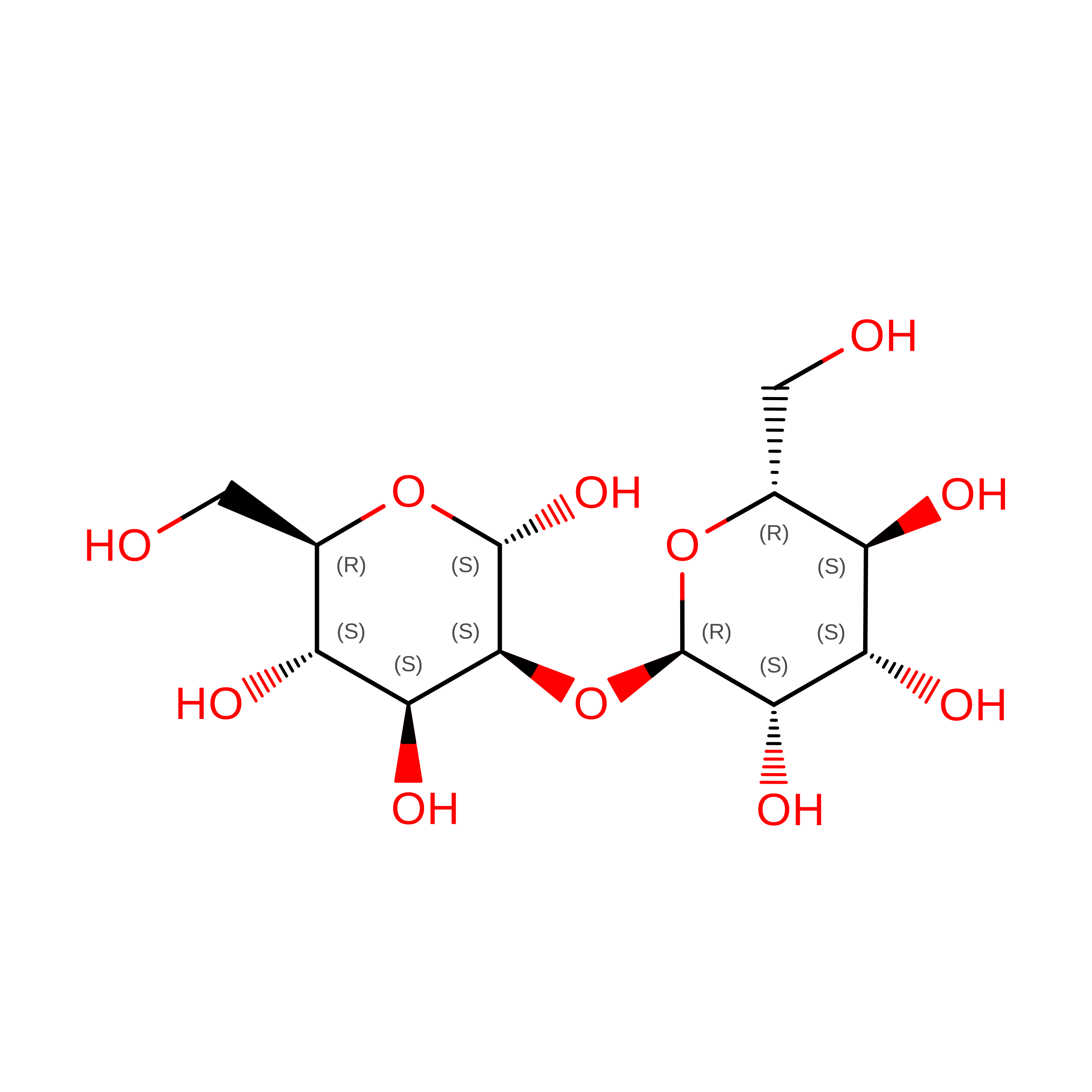
alpha-D-mannopyranose-(1-2)-alpha-D-mannopyranose
C, D
2N/A
Glycosylation Resources
GlyTouCan: G53402KW
GlyCosmos: G53402KW
GlyGen: G53402KW

Biologically Interesting Molecules (External Reference) 

1 Unique

Experimental Data & Validation

Experimental Data

  • Method: X-RAY DIFFRACTION
  • Resolution: 1.80 Å
  • R-Value Free:  0.215 (Depositor), 0.183 (DCC) 
  • R-Value Work:  0.169 (Depositor), 0.191 (DCC) 
  • R-Value Observed: 0.171 (Depositor) 
Space Group: P 1 21 1
Unit Cell:
Length ( Å )Angle ( ˚ )
a = 49.226α = 90
b = 38.281β = 99.69
c = 55.794γ = 90
Software Package:
Software NamePurpose
PHASERphasing
REFMACrefinement
CrystalCleardata collection
HKL-2000data reduction
HKL-2000data scaling

Structure Validation

View Full Validation Report


Entry History 

Deposition Data

Revision History  (Full details and data files)

  • Version 1.0: 2007-07-31
    Type: Initial release
  • Version 1.1: 2011-07-13
    Changes: Version format compliance
  • Version 2.0: 2020-07-29
    Type: Remediation
    Reason: Carbohydrate remediation
    Changes: Advisory, Atomic model, Data collection, Database references, Derived calculations, Structure summary
  • Version 2.1: 2021-10-20
    Changes: Database references, Structure summary
  • Version 2.2: 2023-08-30
    Changes: Data collection, Refinement description
  • Version 2.3: 2024-10-30
    Changes: Structure summary