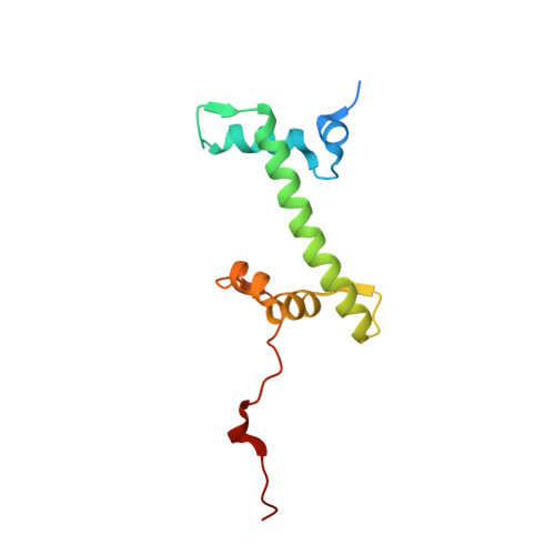
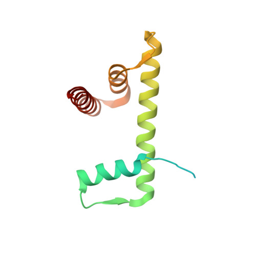
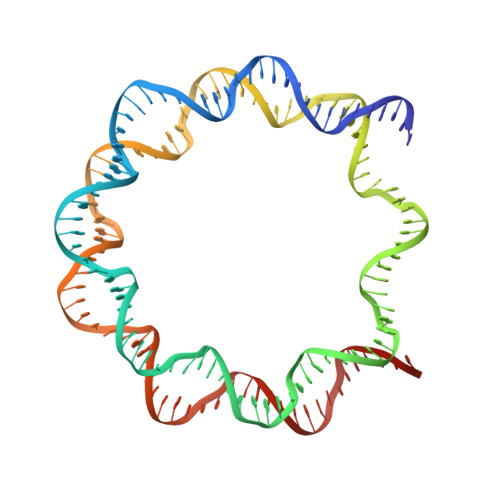
2PYO

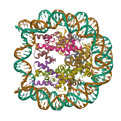
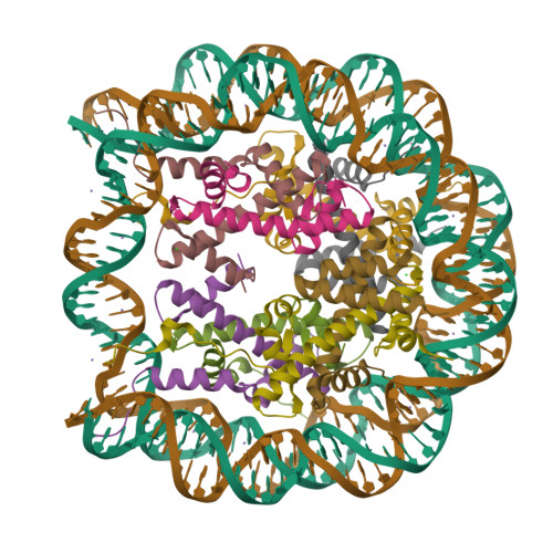
Drosophila nucleosome core


Experimental Data Snapshot

  • Method: X-RAY DIFFRACTION
  • Resolution: 2.43 Å
  • R-Value Free: 0.262 
  • R-Value Work: 0.229 

wwPDB Validation   3D Report Full Report


This is version 1.2 of the entry. See complete history


Literature

Structure of the Drosophila nucleosome core particle highlights evolutionary constraints on the H2A-H2B histone dimer.

Clapier, C.R.Chakravarthy, S.Petosa, C.Fernandez-Tornero, C.Luger, K.Muller, C.W.

(2007) Proteins 71: 1-7

  • DOI: https://doi.org/10.1002/prot.21720
  • Primary Citation of Related Structures:  
    2PYO

  • PubMed Abstract: 

    We determined the 2.45 A crystal structure of the nucleosome core particle from Drosophila melanogaster and compared it to that of Xenopus laevis bound to the identical 147 base-pair DNA fragment derived from human alpha-satellite DNA. Differences between the two structures primarily reflect 16 amino acid substitutions between species, 15 of which are in histones H2A and H2B. Four of these involve histone tail residues, resulting in subtly altered protein-DNA interactions that exemplify the structural plasticity of these tails. Of the 12 substitutions occurring within the histone core regions, five involve small, solvent-exposed residues not involved in intraparticle interactions. The remaining seven involve buried hydrophobic residues, and appear to have coevolved so as to preserve the volume of side chains within the H2A hydrophobic core and H2A-H2B dimer interface. Thus, apart from variations in the histone tails, amino acid substitutions that differentiate Drosophila from Xenopus histones occur in mutually compensatory combinations. This highlights the tight evolutionary constraints exerted on histones since the vertebrate and invertebrate lineages diverged.


  • Organizational Affiliation

    European Molecular Biology Laboratory, Grenoble Outstation, 38042 Grenoble Cedex 9, France.


Macromolecules

Find similar proteins by:  (by identity cutoff)  |  3D Structure
Entity ID: 3
MoleculeChains Sequence LengthOrganismDetailsImage
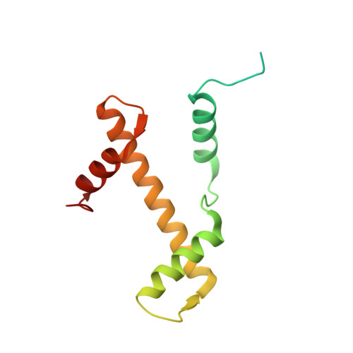
Histone H3C [auth A],
G [auth E]
135Drosophila melanogasterMutation(s): 0 
Gene Names: His3
UniProt
Find proteins for P02299 (Drosophila melanogaster)
Explore P02299 
Go to UniProtKB:  P02299
Entity Groups  
Sequence Clusters30% Identity50% Identity70% Identity90% Identity95% Identity100% Identity
UniProt GroupP02299
Sequence Annotations
Expand
  • Reference Sequence
Find similar proteins by:  (by identity cutoff)  |  3D Structure
Entity ID: 4
MoleculeChains Sequence LengthOrganismDetailsImage
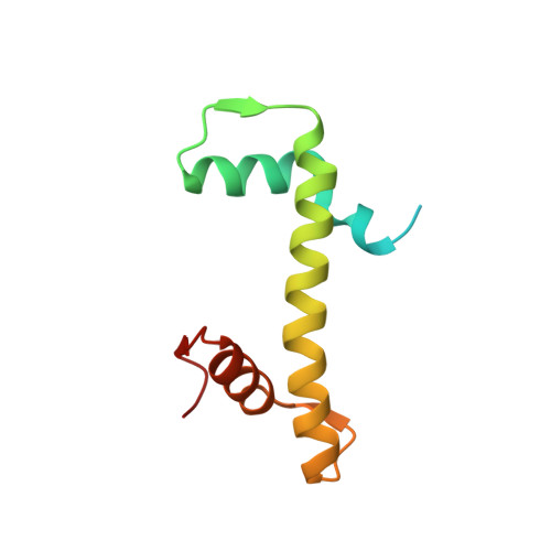
Histone H4D [auth B],
H [auth F]
102Drosophila melanogasterMutation(s): 0 
Gene Names: His4H4
UniProt
Find proteins for P84040 (Drosophila melanogaster)
Explore P84040 
Go to UniProtKB:  P84040
Entity Groups  
Sequence Clusters30% Identity50% Identity70% Identity90% Identity95% Identity100% Identity
UniProt GroupP84040
Sequence Annotations
Expand
  • Reference Sequence
Find similar proteins by:  (by identity cutoff)  |  3D Structure
Entity ID: 5
MoleculeChains Sequence LengthOrganismDetailsImage
Histone H2AE [auth C],
I [auth G]
120Drosophila melanogasterMutation(s): 0 
Gene Names: His2AH2a
UniProt
Find proteins for P84051 (Drosophila melanogaster)
Explore P84051 
Go to UniProtKB:  P84051
Entity Groups  
Sequence Clusters30% Identity50% Identity70% Identity90% Identity95% Identity100% Identity
UniProt GroupP84051
Sequence Annotations
Expand
  • Reference Sequence
Find similar proteins by:  (by identity cutoff)  |  3D Structure
Entity ID: 6
MoleculeChains Sequence LengthOrganismDetailsImage
Histone H2BF [auth D],
J [auth H]
122Drosophila melanogasterMutation(s): 0 
Gene Names: His2B
UniProt
Find proteins for P02283 (Drosophila melanogaster)
Explore P02283 
Go to UniProtKB:  P02283
Entity Groups  
Sequence Clusters30% Identity50% Identity70% Identity90% Identity95% Identity100% Identity
UniProt GroupP02283
Sequence Annotations
Expand
  • Reference Sequence
Find similar nucleic acids by:  (by identity cutoff)  |  3D Structure
Entity ID: 1
MoleculeChains LengthOrganismImage
DNA (147-MER)A [auth I]147Homo sapiens
Sequence Annotations
Expand
  • Reference Sequence
Find similar nucleic acids by:  (by identity cutoff)  |  3D Structure
Entity ID: 2
MoleculeChains LengthOrganismImage
DNA (147-MER)B [auth J]147Homo sapiens
Sequence Annotations
Expand
  • Reference Sequence
Small Molecules
Ligands 2 Unique
IDChains Name / Formula / InChI Key2D Diagram3D Interactions
MN
Query on MN

Download Ideal Coordinates CCD File 
K [auth I]
L [auth I]
M [auth I]
N [auth I]
O [auth I]
K [auth I],
L [auth I],
M [auth I],
N [auth I],
O [auth I],
P [auth I],
Q [auth I],
R [auth J],
S [auth J],
T [auth J],
U [auth J],
V [auth J],
W [auth J],
Z [auth E]
MANGANESE (II) ION
Mn
WAEMQWOKJMHJLA-UHFFFAOYSA-N
CL
Query on CL

Download Ideal Coordinates CCD File 
AA [auth E],
BA [auth H],
X [auth A],
Y [auth D]
CHLORIDE ION
Cl
VEXZGXHMUGYJMC-UHFFFAOYSA-M
Experimental Data & Validation

Experimental Data

  • Method: X-RAY DIFFRACTION
  • Resolution: 2.43 Å
  • R-Value Free: 0.262 
  • R-Value Work: 0.229 
  • Space Group: P 21 21 21
Unit Cell:
Length ( Å )Angle ( ˚ )
a = 106.03α = 90
b = 182.04β = 90
c = 109.42γ = 90
Software Package:
Software NamePurpose
XSCALEdata processing
CNSrefinement
PDB_EXTRACTdata extraction
XDSdata reduction
XSCALEdata scaling
CNSphasing

Structure Validation

View Full Validation Report


Entry History 

Deposition Data

Revision History  (Full details and data files)

  • Version 1.0: 2007-11-06
    Type: Initial release
  • Version 1.1: 2011-07-13
    Changes: Version format compliance
  • Version 1.2: 2024-02-21
    Changes: Data collection, Database references, Derived calculations