2PY2 | pdb_00002py2

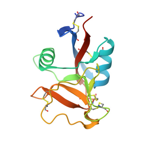
Structure of Herring Type II Antifreeze Protein


Experimental Data Snapshot

  • Method: X-RAY DIFFRACTION
  • Resolution: 1.70 Å
  • R-Value Free: 
    0.240 (Depositor) 
  • R-Value Work: 
    0.209 (Depositor), 0.217 (DCC) 

wwPDB Validation 3D Report Full Report

Validation slider image for 2PY2

This is version 1.3 of the entry. See complete history

Literature

Structure and Evolutionary Origin of Ca-Dependent Herring Type II Antifreeze Protein.

Liu, Y.Li, Z.Lin, Q.Kosinski, J.Seetharaman, J.Bujnicki, J.M.Sivaraman, J.Hew, C.L.

(2007) PLoS One 2: e548-e548

  • DOI: https://doi.org/10.1371/journal.pone.0000548
  • Primary Citation Related Structures: 
    2PY2

  • PubMed Abstract: 

    In order to survive under extremely cold environments, many organisms produce antifreeze proteins (AFPs). AFPs inhibit the growth of ice crystals and protect organisms from freezing damage. Fish AFPs can be classified into five distinct types based on their structures. Here we report the structure of herring AFP (hAFP), a Ca(2+)-dependent fish type II AFP. It exhibits a fold similar to the C-type (Ca(2+)-dependent) lectins with unique ice-binding features. The 1.7 A crystal structure of hAFP with bound Ca(2+) and site-directed mutagenesis reveal an ice-binding site consisting of Thr96, Thr98 and Ca(2+)-coordinating residues Asp94 and Glu99, which initiate hAFP adsorption onto the [10-10] prism plane of the ice lattice. The hAFP-ice interaction is further strengthened by the bound Ca(2+) through the coordination with a water molecule of the ice lattice. This Ca(2+)-coordinated ice-binding mechanism is distinct from previously proposed mechanisms for other AFPs. However, phylogenetic analysis suggests that all type II AFPs evolved from the common ancestor and developed different ice-binding modes. We clarify the evolutionary relationship of type II AFPs to sugar-binding lectins.


  • Organizational Affiliation
    • Department of Biological Sciences, National University of Singapore, Singapore, Singapore.

Macromolecule Content 

  • Total Structure Weight: 92.99 kDa 
  • Atom Count: 6,557 
  • Modeled Residue Count: 762 
  • Deposited Residue Count: 816 
  • Unique protein chains: 1

Macromolecules

Find similar proteins by:|  3D Structure
Entity ID: 1
MoleculeChains  Sequence LengthOrganismDetailsImage
Antifreeze protein type II
A, B, C, D, E
A, B, C, D, E, F
136Clupea harengusMutation(s): 0 
Gene Names: hAFP
UniProt
Find proteins for Q91992 (Clupea harengus)
Explore Q91992 
Go to UniProtKB:  Q91992
Entity Groups
Sequence Clusters30% Identity50% Identity70% Identity90% Identity95% Identity100% Identity
UniProt GroupQ91992
Sequence Annotations
Expand
Reference Sequence

Experimental Data & Validation

Experimental Data

  • Method: X-RAY DIFFRACTION
  • Resolution: 1.70 Å
  • R-Value Free:  0.240 (Depositor) 
  • R-Value Work:  0.209 (Depositor), 0.217 (DCC) 
Space Group: P 21 21 21
Unit Cell:
Length ( Å )Angle ( ˚ )
a = 31.279α = 90
b = 146.415β = 90
c = 192.406γ = 90
Software Package:
Software NamePurpose
HKL-2000data collection
SOLVEphasing
CNSrefinement
HKL-2000data reduction
HKL-2000data scaling

Structure Validation

View Full Validation Report


Entry History 

Deposition Data

Revision History  (Full details and data files)

  • Version 1.0: 2007-06-26
    Type: Initial release
  • Version 1.1: 2008-05-01
    Changes: Version format compliance
  • Version 1.2: 2011-07-13
    Changes: Version format compliance
  • Version 1.3: 2024-11-20
    Changes: Data collection, Database references, Derived calculations, Structure summary