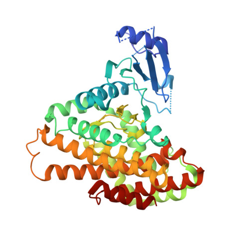
2PUL | pdb_00002pul

Structures of 5-methylthioribose kinase reveal substrate specificity and unusual mode of nucleotide binding


Experimental Data Snapshot

  • Method: X-RAY DIFFRACTION
  • Resolution: 2.00 Å
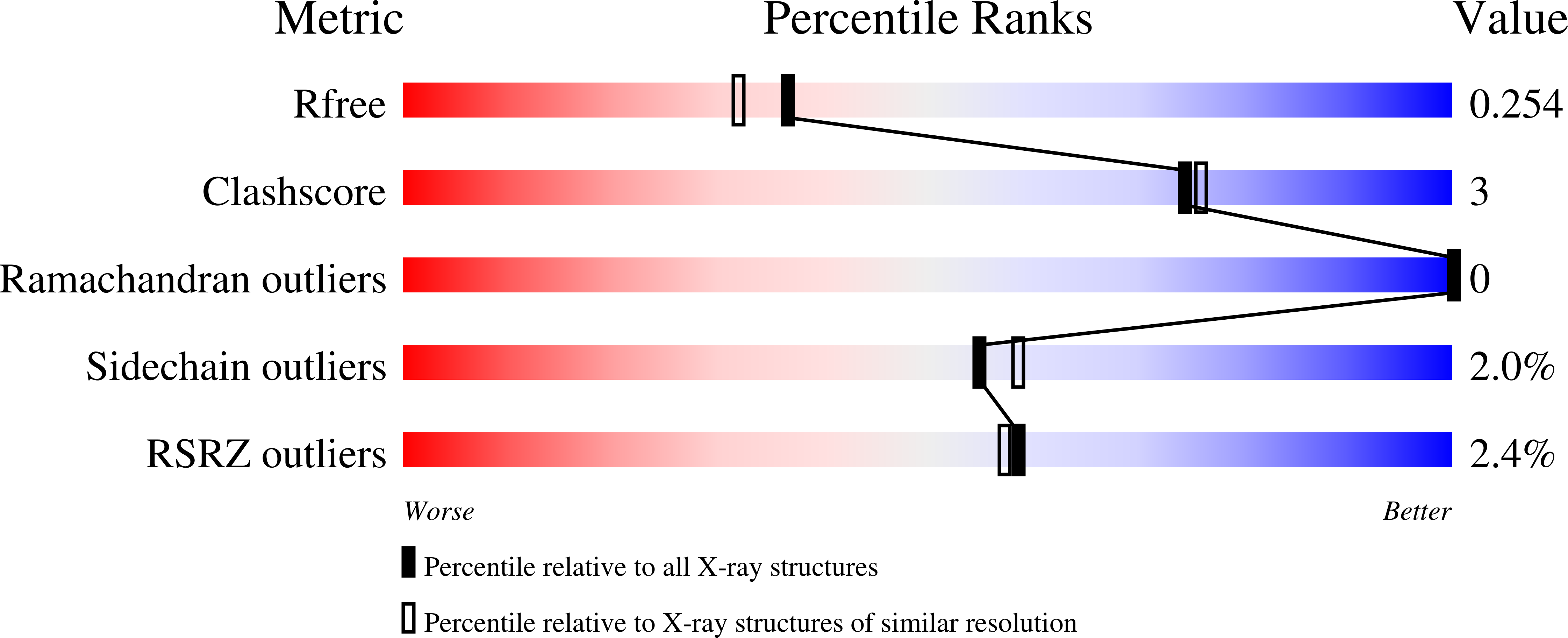
  • R-Value Free: 
    0.240 (Depositor), 0.250 (DCC) 
  • R-Value Work: 
    0.203 (Depositor), 0.220 (DCC) 
  • R-Value Observed: 
    0.205 (Depositor) 

wwPDB Validation   3D Report Full Report


Ligand Structure Quality Assessment 

Created with Raphaël 2.3.0Worse 01 BetterLigand structure goodness of fit to experimental dataBest fitted CPSClick on this verticalbar to view detailsBest fitted ACPClick on this verticalbar to view details

This is version 1.3 of the entry. See complete history


Literature

Structures of 5-methylthioribose kinase reveal substrate specificity and unusual mode of nucleotide binding

Ku, S.-Y.Yip, P.Cornell, K.A.Riscoe, M.K.Behr, J.-B.Guillerm, G.Howell, P.L.

(2007) J Biological Chem 282: 22195-22206

  • DOI: https://doi.org/10.1074/jbc.M611045200
  • Primary Citation of Related Structures:  
    2PU8, 2PUI, 2PUL, 2PUN, 2PUP

  • PubMed Abstract: 

    The methionine salvage pathway is ubiquitous in all organisms, but metabolic variations exist between bacteria and mammals. 5-Methylthioribose (MTR) kinase is a key enzyme in methionine salvage in bacteria and the absence of a mammalian homolog suggests that it is a good target for the design of novel antibiotics. The structures of the apo-form of Bacillus subtilis MTR kinase, as well as its ADP, ADP-PO(4), AMPPCP, and AMPPCP-MTR complexes have been determined. MTR kinase has a bilobal eukaryotic protein kinase fold but exhibits a number of unique features. The protein lacks the DFG motif typically found at the beginning of the activation loop and instead coordinates magnesium via a DXE motif (Asp(250)-Glu(252)). In addition, the glycine-rich loop of the protein, analogous to the "Gly triad" in protein kinases, does not interact extensively with the nucleotide. The MTR substrate-binding site consists of Asp(233) of the catalytic HGD motif, a novel twin arginine motif (Arg(340)/Arg(341)), and a semi-conserved W-loop, which appears to regulate MTR binding specificity. No lobe closure is observed for MTR kinase upon substrate binding. This is probably because the enzyme lacks the lobe closure/inducing interactions between the C-lobe of the protein and the ribosyl moiety of the nucleotide that are typically responsible for lobe closure in protein kinases. The current structures suggest that MTR kinase has a dissociative mechanism.


  • Organizational Affiliation

    Program in Molecular Structure and Function, Research Institute, Hospital for Sick Children, Toronto, Ontario, Canada.


Macromolecules
Find similar proteins by:  (by identity cutoff)  |  3D Structure
Entity ID: 1
MoleculeChains Sequence LengthOrganismDetailsImage
Methylthioribose kinase
A, B
397Bacillus subtilisMutation(s): 0 
Gene Names: mtnKykrT
EC: 2.7.1.100
UniProt
Find proteins for O31663 (Bacillus subtilis (strain 168))
Explore O31663 
Go to UniProtKB:  O31663
Entity Groups  
Sequence Clusters30% Identity50% Identity70% Identity90% Identity95% Identity100% Identity
UniProt GroupO31663
Sequence Annotations
Expand
  • Reference Sequence
Experimental Data & Validation

Experimental Data

  • Method: X-RAY DIFFRACTION
  • Resolution: 2.00 Å
  • R-Value Free:  0.240 (Depositor), 0.250 (DCC) 
  • R-Value Work:  0.203 (Depositor), 0.220 (DCC) 
  • R-Value Observed: 0.205 (Depositor) 
Space Group: P 21 21 2
Unit Cell:
Length ( Å )Angle ( ˚ )
a = 214.15α = 90
b = 83.47β = 90
c = 51.26γ = 90
Software Package:
Software NamePurpose
REFMACrefinement
d*TREKdata scaling
d*TREKdata reduction
CNSphasing

Structure Validation

View Full Validation Report


Ligand Structure Quality Assessment 

Created with Raphaël 2.3.0Worse 01 BetterLigand structure goodness of fit to experimental dataBest fitted CPSClick on this verticalbar to view detailsBest fitted ACPClick on this verticalbar to view details

Entry History 

Deposition Data

  • Released Date: 2007-05-22 
  • Deposition Author(s): Ku, S.-Y.

Revision History  (Full details and data files)

  • Version 1.0: 2007-05-22
    Type: Initial release
  • Version 1.1: 2008-05-01
    Changes: Version format compliance
  • Version 1.2: 2011-07-13
    Changes: Version format compliance
  • Version 1.3: 2024-02-21
    Changes: Data collection, Database references, Derived calculations, Refinement description