2PUH | pdb_00002puh

Crystal Structure of the S112A mutant of a C-C hydrolase, BphD from Burkholderia xenovorans LB400, in complex with its substrate HOPDA


Experimental Data Snapshot

  • Method: X-RAY DIFFRACTION
  • Resolution: 1.82 Å
  • R-Value Free: 
    0.203 (Depositor), 0.215 (DCC) 
  • R-Value Work: 
    0.168 (Depositor), 0.176 (DCC) 
  • R-Value Observed: 
    0.170 (Depositor) 

Starting Model: experimental
View more details

wwPDB Validation 3D Report Full Report

Validation slider image for 2PUH

Ligand Structure Quality Assessment 


This is version 1.5 of the entry. See complete history

Literature

The Tautomeric Half-reaction of BphD, a C-C Bond Hydrolase: KINETIC AND STRUCTURAL EVIDENCE SUPPORTING A KEY ROLE FOR HISTIDINE 265 OF THE CATALYTIC TRIAD.

Horsman, G.P.Bhowmik, S.Seah, S.Y.Kumar, P.Bolin, J.T.Eltis, L.D.

(2007) J Biological Chem 282: 19894-19904

  • DOI: https://doi.org/10.1074/jbc.M702237200
  • Primary Citation Related Structures: 
    2PU5, 2PU7, 2PUH, 2PUJ, 2RI6

  • PubMed Abstract: 

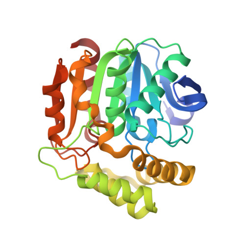
    BphD of Burkholderia xenovorans LB400 catalyzes an unusual C-C bond hydrolysis of 2-hydroxy-6-oxo-6-phenylhexa-2,4-dienoic acid (HOPDA) to afford benzoic acid and 2-hydroxy-2,4-pentadienoic acid (HPD). An enol-keto tautomerization has been proposed to precede hydrolysis via a gem-diol intermediate. The role of the canonical catalytic triad (Ser-112, His-265, Asp-237) in mediating these two half-reactions remains unclear. We previously reported that the BphD-catalyzed hydrolysis of HOPDA (lambda(max) is 434 nm for the free enolate) proceeds via an unidentified intermediate with a red-shifted absorption spectrum (lambda(max) is 492 nm) (Horsman, G. P., Ke, J., Dai, S., Seah, S. Y. K., Bolin, J. T., and Eltis, L. D. (2006) Biochemistry 45, 11071-11086). Here we demonstrate that the S112A variant generates and traps a similar intermediate (lambda(max) is 506 nm) with a similar rate, 1/tau approximately 500 s(-1). The crystal structure of the S112A:HOPDA complex at 1.8-A resolution identified this intermediate as the keto tautomer, (E)-2,6-dioxo-6-phenyl-hex-3-enoate. This keto tautomer did not accumulate in either the H265A or the S112A/H265A double variants, indicating that His-265 catalyzes tautomerization. Consistent with this role, the wild type and S112A enzymes catalyzed tautomerization of the product HPD, whereas H265A variants did not. This study thus identifies a keto intermediate, and demonstrates that the catalytic triad histidine catalyzes the tautomerization half-reaction, expanding the role of this residue from its purely hydrolytic function in other serine hydrolases. Finally, the S112A:HOPDA crystal structure is more consistent with hydrolysis occurring via an acyl-enzyme intermediate than a gem-diol intermediate as solvent molecules have poor access to C6, and the closest ordered water is 7 A away.


  • Organizational Affiliation
    • Department of Biochemistry, University of British Columbia, Vancouver, British Columbia V6T 1Z3, Canada.

Macromolecule Content 

  • Total Structure Weight: 32.39 kDa 
  • Atom Count: 2,388 
  • Modeled Residue Count: 283 
  • Deposited Residue Count: 286 
  • Unique protein chains: 1

Macromolecules

Find similar proteins by:|  3D Structure
Entity ID: 1
MoleculeChains  Sequence LengthOrganismDetailsImage
2-hydroxy-6-oxo-6-phenylhexa-2,4-dienoate hydrolase286Paraburkholderia xenovoransMutation(s): 1 
Gene Names: bphD
EC: 3.7.1 (PDB Primary Data), 3.7.1.8 (UniProt)
UniProt
Find proteins for P47229 (Paraburkholderia xenovorans (strain LB400))
Explore P47229 
Go to UniProtKB:  P47229
Entity Groups
Sequence Clusters30% Identity50% Identity70% Identity90% Identity95% Identity100% Identity
UniProt GroupP47229
Sequence Annotations
Expand
Reference Sequence

Experimental Data & Validation

Experimental Data

  • Method: X-RAY DIFFRACTION
  • Resolution: 1.82 Å
  • R-Value Free:  0.203 (Depositor), 0.215 (DCC) 
  • R-Value Work:  0.168 (Depositor), 0.176 (DCC) 
  • R-Value Observed: 0.170 (Depositor) 
Space Group: I 41 2 2
Unit Cell:
Length ( Å )Angle ( ˚ )
a = 116.808α = 90
b = 116.808β = 90
c = 87.589γ = 90
Software Package:
Software NamePurpose
REFMACrefinement
PDB_EXTRACTdata extraction
HKL-2000data reduction
HKL-2000data scaling
MOLREPphasing

Structure Validation

View Full Validation Report


Ligand Structure Quality Assessment 


Entry History 

Deposition Data

Revision History  (Full details and data files)

  • Version 1.0: 2007-05-22
    Type: Initial release
  • Version 1.1: 2008-05-01
    Changes: Version format compliance
  • Version 1.2: 2011-07-13
    Changes: Derived calculations, Version format compliance
  • Version 1.3: 2017-10-18
    Changes: Refinement description
  • Version 1.4: 2021-10-20
    Changes: Database references, Derived calculations
  • Version 1.5: 2023-08-30
    Changes: Data collection, Refinement description