X-ray crystallographic characterization of the Co(II)-substituted Tris-bound form of the aminopeptidase from Aeromonas proteolytica.
Munih, P., Moulin, A., Stamper, C.C., Bennett, B., Ringe, D., Petsko, G.A., Holz, R.C.(2007) J Inorg Biochem 101: 1099-1107
- PubMed: 17574677 Search on PubMed
- DOI: https://doi.org/10.1016/j.jinorgbio.2007.03.010
- Primary Citation Related Structures:
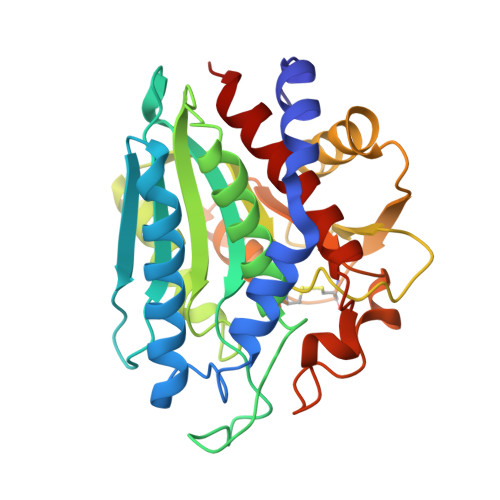
2PRQ - PubMed Abstract:
The X-ray crystal structure of the Co(II)-loaded form of the aminopeptidase from Aeromonas proteolytica ([CoCo(AAP)]) was solved to 2.2A resolution. [CoCo(AAP)] folds into an alpha/beta globular domain with a twisted beta-sheet hydrophobic core sandwiched between alpha-helices, identical to [ZnZn(AAP)]. Co(II) binding to AAP does not introduce any major conformational changes to the overall protein structure and the amino acid residues ligated to the dicobalt(II) cluster in [CoCo(AAP)] are the same as those in the native Zn(II)-loaded structure with only minor perturbations in bond lengths. The Co(II)-Co(II) distance is 3.3A. Tris(hydroxymethyl)aminomethane (Tris) coordinates to the dinuclear Co(II) active site of AAP with one of the Tris hydroxyl oxygen atoms (O4) forming a single oxygen atom bridge between the two Co(II) ions. This is the only Tris atom coordinated to the metals with Co1-O and Co2-O bonds distances of 2.2 and 1.9A, respectively. Each of the Co(II) ions resides in a distorted trigonal bipyramidal geometry. This important structure bridges the gap between previous structural and spectroscopic studies performed on AAP and is discussed in this context.
- Program in Biophysics and Structural Biology, Rosenstiel Basic Medical Sciences Research Center, Brandeis University, 415 South Street, Waltham, MA 02254, United States.
Organizational Affiliation: