2PRE | pdb_00002pre

Crystal structure of plant cysteine protease Ervatamin-C complexed with irreversible inhibitor E-64 at 2.7 A resolution


Experimental Data Snapshot

  • Method: X-RAY DIFFRACTION
  • Resolution: 2.70 Å
  • R-Value Free: 
    0.234 (Depositor), 0.220 (DCC) 
  • R-Value Work: 
    0.197 (Depositor), 0.189 (DCC) 
  • R-Value Observed: 
    0.197 (Depositor) 

Starting Model: experimental
View more details

wwPDB Validation 3D Report Full Report

Validation slider image for 2PRE

Ligand Structure Quality Assessment 


This is version 1.3 of the entry. See complete history

Literature

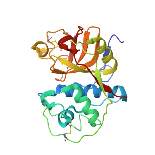
Structural insights into the substrate specificity and activity of ervatamins, the papain-like cysteine proteases from a tropical plant, Ervatamia coronaria.

Ghosh, R.Chakraborty, S.Chakrabarti, C.Dattagupta, J.K.Biswas, S.

(2008) FEBS J 275: 421-434

  • DOI: https://doi.org/10.1111/j.1742-4658.2007.06211.x
  • Primary Citation Related Structures: 
    2PRE, 3BCN

  • PubMed Abstract: 

    Multiple proteases of the same family are quite often present in the same species in biological systems. These multiple proteases, despite having high homology in their primary and tertiary structures, show deviations in properties such as stability, activity, and specificity. It is of interest, therefore, to compare the structures of these multiple proteases in a single species to identify the structural changes, if any, that may be responsible for such deviations. Ervatamin-A, ervatamin-B and ervatamin-C are three such papain-like cysteine proteases found in the latex of the tropical plant Ervatamia coronaria, and are known not only for their high stability over a wide range of temperature and pH, but also for variations in activity and specificity among themselves and among other members of the family. Here we report the crystal structures of ervatamin-A and ervatamin-C, complexed with an irreversible inhibitor 1-[l-N-(trans-epoxysuccinyl)leucyl]amino-4-guanidinobutane (E-64), together with enzyme kinetics and molecular dynamic simulation studies. A comparison of these results with the earlier structures helps in a correlation of the structural features with the corresponding functional properties. The specificity constants (k(cat)/K(m)) for the ervatamins indicate that all of these enzymes have specificity for a branched hydrophobic residue at the P2 position of the peptide substrates, with different degrees of efficiency. A single amino acid change, as compared to ervatamin-C, in the S2 pocket of ervatamin-A (Ala67-->Tyr) results in a 57-fold increase in its k(cat)/K(m) value for a substrate having a Val at the P2 position. Our studies indicate a higher enzymatic activity of ervatamin-A, which has been subsequently explained at the molecular level from the three-dimensional structure of the enzyme and in the context of its helix polarizibility and active site plasticity.


  • Organizational Affiliation
    • Crystallography and Molecular Biology Division, Saha Institute of Nuclear Physics, Kolkata, India.

Macromolecule Content 

  • Total Structure Weight: 47.29 kDa 
  • Atom Count: 3,480 
  • Modeled Residue Count: 416 
  • Deposited Residue Count: 416 
  • Unique protein chains: 1

Macromolecules

Find similar proteins by:|  3D Structure
Entity ID: 1
MoleculeChains  Sequence LengthOrganismDetailsImage
Ervatamin-C
A, B
208Tabernaemontana divaricataMutation(s): 0 
EC: 3.4.22
UniProt
Find proteins for A8DS38 (Tabernaemontana divaricata)
Explore A8DS38 
Go to UniProtKB:  A8DS38
Entity Groups
Sequence Clusters30% Identity50% Identity70% Identity90% Identity95% Identity100% Identity
UniProt GroupA8DS38
Sequence Annotations
Expand
Reference Sequence

Small Molecules

Ligands 2 Unique
IDChains Name / Formula / InChI Key2D Diagram3D Interactions
E64

Query on E64


Download:Ideal Coordinates CCD File
F [auth A],
J [auth B]
N-[N-[1-HYDROXYCARBOXYETHYL-CARBONYL]LEUCYLAMINO-BUTYL]-GUANIDINE
C15 H30 N5 O5
QPQNJAXBPHVASB-QWRGUYRKSA-O
SO4

Query on SO4


Download:Ideal Coordinates CCD File
C [auth A]
D [auth A]
E [auth A]
G [auth B]
H [auth B]
C [auth A],
D [auth A],
E [auth A],
G [auth B],
H [auth B],
I [auth B]
SULFATE ION
O4 S
QAOWNCQODCNURD-UHFFFAOYSA-L

Experimental Data & Validation

Experimental Data

  • Method: X-RAY DIFFRACTION
  • Resolution: 2.70 Å
  • R-Value Free:  0.234 (Depositor), 0.220 (DCC) 
  • R-Value Work:  0.197 (Depositor), 0.189 (DCC) 
  • R-Value Observed: 0.197 (Depositor) 
Space Group: P 21 21 21
Unit Cell:
Length ( Å )Angle ( ˚ )
a = 43.37α = 90
b = 81.46β = 90
c = 131.95γ = 90
Software Package:
Software NamePurpose
CNSrefinement
MAR345dtbdata collection
AUTOMARdata reduction
AMoREphasing

Structure Validation

View Full Validation Report


Ligand Structure Quality Assessment 


Entry History 

Deposition Data

Revision History  (Full details and data files)

  • Version 1.0: 2008-01-22
    Type: Initial release
  • Version 1.1: 2011-07-13
    Changes: Version format compliance
  • Version 1.2: 2023-08-30
    Changes: Data collection, Database references, Derived calculations, Refinement description
  • Version 1.3: 2024-10-16
    Changes: Structure summary