2PQS | pdb_00002pqs

Crystal Structure of the Bovine Lactadherin C2 Domain


Experimental Data Snapshot

  • Method: X-RAY DIFFRACTION
  • Resolution: 2.40 Å
  • R-Value Free: 
    0.273 (Depositor), 0.273 (DCC) 
  • R-Value Work: 
    0.235 (Depositor), 0.235 (DCC) 
  • R-Value Observed: 
    0.235 (Depositor) 

wwPDB Validation 3D Report Full Report

Validation slider image for 2PQS

This is version 1.2 of the entry. See complete history

Literature

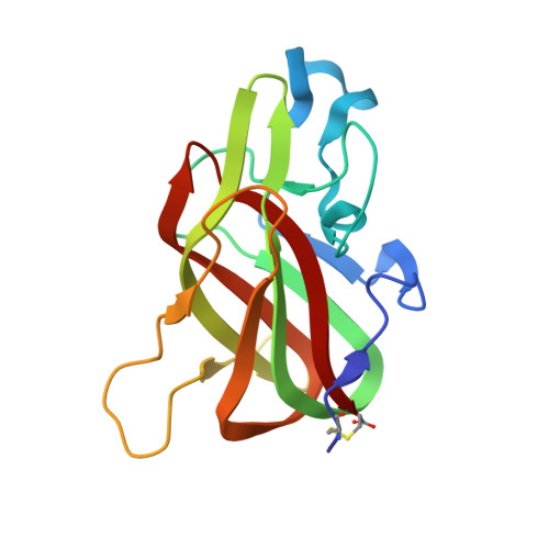
Crystal structure of the bovine lactadherin C2 domain, a membrane binding motif, shows similarity to the C2 domains of factor V and factor VIII.

Lin, L.Huai, Q.Huang, M.Furie, B.Furie, B.C.

(2007) J Mol Biology 371: 717-724

  • DOI: https://doi.org/10.1016/j.jmb.2007.05.054
  • Primary Citation Related Structures: 
    2PQS

  • PubMed Abstract: 

    Lactadherin, a glycoprotein secreted by a variety of cell types, contains two EGF domains and two C domains with sequence homology to the C domains of blood coagulation proteins factor V and factor VIII. Like these proteins, lactadherin binds to phosphatidylserine (PS)-containing membranes with high affinity. We determined the crystal structure of the bovine lactadherin C2 domain (residues 1 to 158) at 2.4 A. The lactadherin C2 structure is similar to the C2 domains of factors V and VIII (rmsd of C(alpha) atoms of 0.9 A and 1.2 A, and sequence identities of 43% and 38%, respectively). The lactadherin C2 domain has a discoidin-like fold containing two beta-sheets of five and three antiparallel beta-strands packed against one another. The N and C termini are linked by a disulfide bridge between Cys1 and Cys158. One beta-turn and two loops containing solvent-exposed hydrophobic residues extend from the C2 domain beta-sandwich core. In analogy with the C2 domains of factors V and VIII, some or all of these solvent-exposed hydrophobic residues, Trp26, Leu28, Phe31, and Phe81, likely participate in membrane binding. The C2 domain of lactadherin may serve as a marker of cell surface phosphatidylserine exposure and may have potential as a unique anti-thrombotic agent.


  • Organizational Affiliation
    • Division of Hemostasis and Thrombosis, Beth Israel Deaconess Medical Center and Department of Medicine, Harvard Medical School, Boston, MA 02215, USA.

Macromolecule Content 

  • Total Structure Weight: 72.38 kDa 
  • Atom Count: 5,219 
  • Modeled Residue Count: 636 
  • Deposited Residue Count: 636 
  • Unique protein chains: 1

Macromolecules

Find similar proteins by:|  3D Structure
Entity ID: 1
MoleculeChains  Sequence LengthOrganismDetailsImage
Lactadherin
A, B, C, D
159Bos taurusMutation(s): 0 
Gene Names: MFGE8
UniProt
Find proteins for Q95114 (Bos taurus)
Explore Q95114 
Go to UniProtKB:  Q95114
Entity Groups
Sequence Clusters30% Identity50% Identity70% Identity90% Identity95% Identity100% Identity
UniProt GroupQ95114
Sequence Annotations
Expand
Reference Sequence

Experimental Data & Validation

Experimental Data

  • Method: X-RAY DIFFRACTION
  • Resolution: 2.40 Å
  • R-Value Free:  0.273 (Depositor), 0.273 (DCC) 
  • R-Value Work:  0.235 (Depositor), 0.235 (DCC) 
  • R-Value Observed: 0.235 (Depositor) 
Space Group: P 21 21 2
Unit Cell:
Length ( Å )Angle ( ˚ )
a = 108.123α = 90
b = 107.788β = 90
c = 82.754γ = 90
Software Package:
Software NamePurpose
CNSrefinement
HKL-2000data collection
HKL-2000data reduction
HKL-2000data scaling
AMoREphasing

Structure Validation

View Full Validation Report


Entry History 

Deposition Data

Revision History  (Full details and data files)

  • Version 1.0: 2007-08-07
    Type: Initial release
  • Version 1.1: 2011-07-13
    Changes: Version format compliance
  • Version 1.2: 2024-11-20
    Changes: Data collection, Database references, Structure summary