


Crystal Structure of thioredoxin-2
Ye, J., Chou, S., Fuselier, J., Li, J., Beckwith, J., Rapoport, T.To be published.
Experimental Data Snapshot
wwPDB Validation 3D Report Full Report

Entity ID: 1 | |||||
|---|---|---|---|---|---|
| Molecule | Chains | Sequence Length | Organism | Details | Image |
| thioredoxin-2 | 155 | Rhodobacter capsulatus | Mutation(s): 0 Gene Names: thioredoxin EC: 1.8.1.8 | 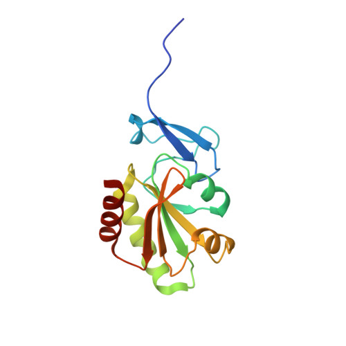 | |
| Ligands 1 Unique | |||||
|---|---|---|---|---|---|
| ID | Chains | Name / Formula / InChI Key | 2D Diagram | 3D Interactions | |
| ZN Download:Ideal Coordinates CCD File | C [auth A], D [auth B], E [auth B] | ZINC ION Zn PTFCDOFLOPIGGS-UHFFFAOYSA-N |  | ||
| Length ( Å ) | Angle ( ˚ ) |
|---|---|
| a = 83.784 | α = 90 |
| b = 68.184 | β = 112.04 |
| c = 64.124 | γ = 90 |
| Software Name | Purpose |
|---|---|
| HKL-3000 | data collection |
| SHELXS | phasing |
| CNS | refinement |
| HKL-3000 | data reduction |
| HKL-3000 | data scaling |


