2PML | pdb_00002pml

Crystal structure of PfPK7 in complex with an ATP analogue


Experimental Data Snapshot

  • Method: X-RAY DIFFRACTION
  • Resolution: 2.60 Å
  • R-Value Free: 
    0.327 (Depositor), 0.312 (DCC) 
  • R-Value Work: 
    0.258 (Depositor), 0.248 (DCC) 
  • R-Value Observed: 
    0.261 (Depositor) 

wwPDB Validation 3D Report Full Report

Validation slider image for 2PML

Ligand Structure Quality Assessment 


This is version 1.2 of the entry. See complete history

Literature

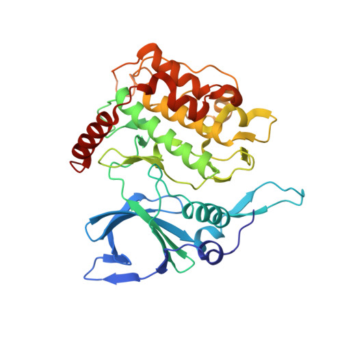
Structures of PfPK7 an atypical protein kinase from P. falciparum identify a novel activation motif and leads for inhibitor design

Merckx, A.Echalier, A.Langford, K.Sicard, A.Langsley, G.Joore, J.Doerig, C.Noble, M.Endicott, J.

To be published.

Macromolecule Content 

  • Total Structure Weight: 42 kDa 
  • Atom Count: 2,927 
  • Modeled Residue Count: 340 
  • Deposited Residue Count: 348 
  • Unique protein chains: 1

Macromolecules

Find similar proteins by:|  3D Structure
Entity ID: 1
MoleculeChains  Sequence LengthOrganismDetailsImage
Ser/Thr protein kinaseA [auth X]348Plasmodium falciparumMutation(s): 0 
Gene Names: PFB0605w
EC: 2.7.12.2
UniProt
Find proteins for O96214 (Plasmodium falciparum)
Explore O96214 
Go to UniProtKB:  O96214
Entity Groups
Sequence Clusters30% Identity50% Identity70% Identity90% Identity95% Identity100% Identity
UniProt GroupO96214
Sequence Annotations
Expand
Reference Sequence

Small Molecules

Ligands 2 Unique
IDChains Name / Formula / InChI Key2D Diagram3D Interactions
ANP

Query on ANP


Download:Ideal Coordinates CCD File
C [auth X]PHOSPHOAMINOPHOSPHONIC ACID-ADENYLATE ESTER
C10 H17 N6 O12 P3
PVKSNHVPLWYQGJ-KQYNXXCUSA-N
MN

Query on MN


Download:Ideal Coordinates CCD File
B [auth X]MANGANESE (II) ION
Mn
WAEMQWOKJMHJLA-UHFFFAOYSA-N

Experimental Data & Validation

Experimental Data

  • Method: X-RAY DIFFRACTION
  • Resolution: 2.60 Å
  • R-Value Free:  0.327 (Depositor), 0.312 (DCC) 
  • R-Value Work:  0.258 (Depositor), 0.248 (DCC) 
  • R-Value Observed: 0.261 (Depositor) 
Space Group: C 2 2 21
Unit Cell:
Length ( Å )Angle ( ˚ )
a = 72.554α = 90
b = 81.795β = 90
c = 138.273γ = 90
Software Package:
Software NamePurpose
REFMACrefinement
DNAdata collection
MOSFLMdata reduction
SCALAdata scaling
SHELXSphasing

Structure Validation

View Full Validation Report


Ligand Structure Quality Assessment 


Entry History 

Deposition Data

Revision History  (Full details and data files)

  • Version 1.0: 2008-01-22
    Type: Initial release
  • Version 1.1: 2011-07-13
    Changes: Advisory, Version format compliance
  • Version 1.2: 2024-02-21
    Changes: Data collection, Database references, Derived calculations