2PLV | pdb_00002plv

STRUCTURAL FACTORS THAT CONTROL CONFORMATIONAL TRANSITIONS AND SEROTYPE SPECIFICITY IN TYPE 3 POLIOVIRUS


Experimental Data Snapshot

  • Method: X-RAY DIFFRACTION
  • Resolution: 2.88 Å
  • R-Value Work: 
    0.200 (Depositor) 

wwPDB Validation 3D Report Full Report

Validation slider image for 2PLV

This is version 2.1 of the entry. See complete history

Literature

Structural factors that control conformational transitions and serotype specificity in type 3 poliovirus

Filman, D.J.Syed, R.Chow, M.Macadam, A.J.Minor, P.D.Hogle, J.M.

(1989) EMBO J 8: 1567-1579

  • DOI: https://doi.org/10.1002/j.1460-2075.1989.tb03541.x
  • Primary Citation Related Structures: 
    2PLV

  • PubMed Abstract: 

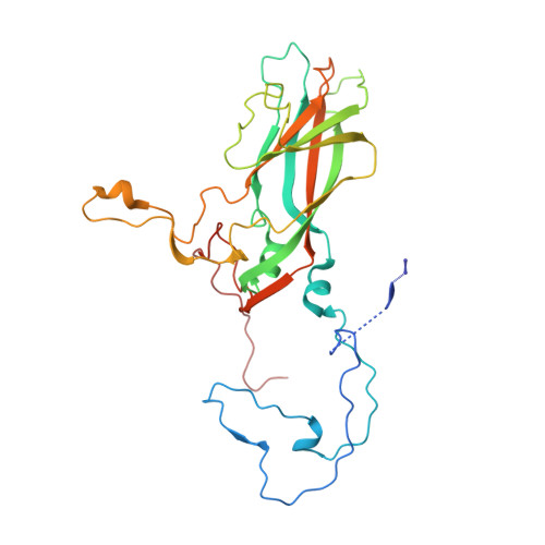
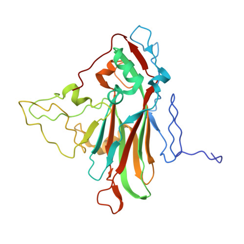
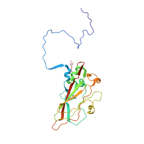
    The three-dimensional structure of the Sabin strain of type 3 poliovirus has been determined at 2.4 A resolution. Significant structural differences with the Mahoney strain of type 1 poliovirus are confined to loops and terminal extensions of the capsid proteins, occur in all of the major antigenic sites of the virion and typically involve insertions, deletions or the replacement of prolines. Several newly identified components of the structure participate in assembly-dependent interactions which are relevant to the biologically important processes of viral assembly and uncoating. These include two sites of lipid substitution, two putative nucleotides and a beta sheet formed by the N-termini of capsid proteins VP4 and VP1. The structure provides an explanation for the temperature sensitive phenotype of the P3/Sabin strain. Amino acids that regulate temperature sensitivity in type 3 poliovirus are located in the interfaces between promoters, in the binding site for a lipid substituent and in an assembly-dependent extended beta sheet that stabilizes the association of pentamers. Several lines of evidence indicate that these structural components also control conformational transitions at various stages of the viral life cycle.


  • Organizational Affiliation
    • Department of Molecular Biology, Research Institute of Scripps Clinic, La Jolla, CA 92037.

Macromolecule Content 

  • Total Structure Weight: 97.92 kDa 
  • Atom Count: 7,162 
  • Modeled Residue Count: 854 
  • Deposited Residue Count: 880 
  • Unique protein chains: 4

Macromolecules

Find similar proteins by:|  3D Structure
Entity ID: 1
MoleculeChains  Sequence LengthOrganismDetailsImage
HUMAN POLIOVIRUS TYPE 1 (SUBUNIT VP1)A [auth 1]302Human poliovirus 1 MahoneyMutation(s): 0 
UniProt
Find proteins for P03300 (Poliovirus type 1 (strain Mahoney))
Explore P03300 
Go to UniProtKB:  P03300
Entity Groups
Sequence Clusters30% Identity50% Identity70% Identity90% Identity95% Identity100% Identity
UniProt GroupP03300
Sequence Annotations
Expand
Reference Sequence
Find similar proteins by:|  3D Structure
Entity ID: 2
MoleculeChains  Sequence LengthOrganismDetailsImage
HUMAN POLIOVIRUS TYPE 1 (SUBUNIT VP2)B [auth 2]272Human poliovirus 1 MahoneyMutation(s): 0 
UniProt
Find proteins for P03300 (Poliovirus type 1 (strain Mahoney))
Explore P03300 
Go to UniProtKB:  P03300
Entity Groups
Sequence Clusters30% Identity50% Identity70% Identity90% Identity95% Identity100% Identity
UniProt GroupP03300
Sequence Annotations
Expand
Reference Sequence
Find similar proteins by:|  3D Structure
Entity ID: 3
MoleculeChains  Sequence LengthOrganismDetailsImage
HUMAN POLIOVIRUS TYPE 1 (SUBUNIT VP3)C [auth 3]238Human poliovirus 1 MahoneyMutation(s): 0 
UniProt
Find proteins for P03300 (Poliovirus type 1 (strain Mahoney))
Explore P03300 
Go to UniProtKB:  P03300
Entity Groups
Sequence Clusters30% Identity50% Identity70% Identity90% Identity95% Identity100% Identity
UniProt GroupP03300
Sequence Annotations
Expand
Reference Sequence
Find similar proteins by:|  3D Structure
Entity ID: 4
MoleculeChains  Sequence LengthOrganismDetailsImage
HUMAN POLIOVIRUS TYPE 1 (SUBUNIT VP4)D [auth 4]68Human poliovirus 1 MahoneyMutation(s): 0 
UniProt
Find proteins for P03300 (Poliovirus type 1 (strain Mahoney))
Explore P03300 
Go to UniProtKB:  P03300
Entity Groups
Sequence Clusters30% Identity50% Identity70% Identity90% Identity95% Identity100% Identity
UniProt GroupP03300
Sequence Annotations
Expand
Reference Sequence

Small Molecules

Ligands 2 Unique
IDChains Name / Formula / InChI Key2D Diagram3D Interactions
SPH

Query on SPH


Download:Ideal Coordinates CCD File
E [auth 1]SPHINGOSINE
C18 H37 N O2
WWUZIQQURGPMPG-MSOLQXFVSA-N
MYR

Query on MYR


Download:Ideal Coordinates CCD File
F [auth 4]MYRISTIC ACID
C14 H28 O2
TUNFSRHWOTWDNC-UHFFFAOYSA-N

Experimental Data & Validation

Experimental Data

  • Method: X-RAY DIFFRACTION
  • Resolution: 2.88 Å
  • R-Value Work:  0.200 (Depositor) 
Space Group: P 21 21 2
Unit Cell:
Length ( Å )Angle ( ˚ )
a = 322.94α = 90
b = 358.04β = 90
c = 380.15γ = 90
Software Package:
Software NamePurpose
REAL-SPACErefinement

Structure Validation

View Full Validation Report


Entry History 

Deposition Data

Revision History  (Full details and data files)

  • Version 1.0: 1989-10-17
    Type: Initial release
  • Version 1.1: 2008-03-24
    Changes: Version format compliance
  • Version 1.2: 2011-07-13
    Changes: Source and taxonomy, Version format compliance
  • Version 1.3: 2017-11-29
    Changes: Derived calculations, Other
  • Version 2.0: 2023-04-19
    Type: Remediation
    Changes: Advisory, Atomic model, Data collection, Database references, Derived calculations, Other, Refinement description
  • Version 2.1: 2024-11-20
    Changes: Data collection, Structure summary