2PLB | pdb_00002plb

D(GTATACC) under hydrostatic pressure of 1.39 GPa


Experimental Data Snapshot

  • Method: X-RAY DIFFRACTION
  • Resolution: 1.60 Å
  • R-Value Free: 
    0.222 (Depositor), 0.226 (DCC) 
  • R-Value Work: 
    0.181 (Depositor), 0.217 (DCC) 
  • R-Value Observed: 
    0.187 (Depositor) 

Starting Model: experimental
View more details

wwPDB Validation 3D Report Full Report

Validation slider image for 2PLB

Ligand Structure Quality Assessment 


This is version 1.3 of the entry. See complete history

Literature

Adaptation of the base-paired double-helix molecular architecture to extreme pressure.

Girard, E.Prange, T.Dhaussy, A.C.Migianu-Griffoni, E.Lecouvey, M.Chervin, J.C.Mezouar, M.Kahn, R.Fourme, R.

(2007) Nucleic Acids Res 35: 4800-4808

  • DOI: https://doi.org/10.1093/nar/gkm511
  • Primary Citation Related Structures: 
    2PKV, 2PL4, 2PL8, 2PLB

  • PubMed Abstract: 

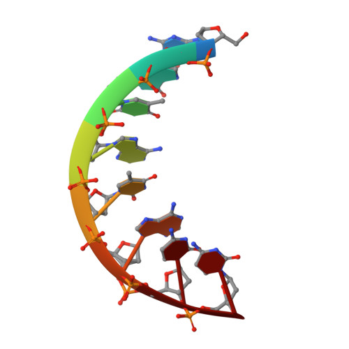
    The behaviour of the d(GGTATACC) oligonucleotide has been investigated by X-ray crystallography at 295 K in the range from ambient pressure to 2 GPa (approximately 20,000 atm). Four 3D-structures of the A-DNA form (at ambient pressure, 0.55, 1.09 and 1.39 GPa) were refined at 1.60 or 1.65 A resolution. In addition to the diffraction pattern of the A-form, the broad meridional streaks previously explained by occluded B-DNA octamers within the channels of the crystalline A-form matrix were observed up to at least 2 GPa. This work highlights an important property of nucleic acids, their capability to withstand very high pressures, while keeping in such conditions a nearly invariant geometry of base pairs that store and carry genetic information. The double-helix base-paired architecture behaves as a molecular spring, which makes it especially adapted to very harsh conditions. These features may have contributed to the emergence of a RNA World at prebiotic stage.


  • Organizational Affiliation
    • Synchrotron-SOLEIL, L'Orme des Merisiers, Saint-Aubin, BP 48, 91192 Gif-sur-Yvette Cedex, France. eric.girard@synchrotron-soleil.fr

Macromolecule Content 

  • Total Structure Weight: 5.06 kDa 
  • Atom Count: 435 
  • Modeled Residue Count: 16 
  • Deposited Residue Count: 16 
  • Unique nucleic acid chains: 1

Macromolecules

Find similar nucleic acids by:  Sequence
Entity ID: 1
MoleculeChains LengthOrganismImage
5'-D(*DGP*DGP*DTP*DAP*DTP*DAP*DCP*DC)-3'
A, B
8N/A
Sequence Annotations
Expand
Reference Sequence

Small Molecules

Ligands 1 Unique
IDChains Name / Formula / InChI Key2D Diagram3D Interactions
SPM

Query on SPM


Download:Ideal Coordinates CCD File
C [auth B]SPERMINE
C10 H26 N4
PFNFFQXMRSDOHW-UHFFFAOYSA-N

Experimental Data & Validation

Experimental Data

  • Method: X-RAY DIFFRACTION
  • Resolution: 1.60 Å
  • R-Value Free:  0.222 (Depositor), 0.226 (DCC) 
  • R-Value Work:  0.181 (Depositor), 0.217 (DCC) 
  • R-Value Observed: 0.187 (Depositor) 
Space Group: P 61
Unit Cell:
Length ( Å )Angle ( ˚ )
a = 42.83α = 90
b = 42.83β = 90
c = 40.3γ = 120
Software Package:
Software NamePurpose
SHELXmodel building
SHELXL-97refinement
AUTOMARdata collection
XDSdata reduction
SCALAdata scaling

Structure Validation

View Full Validation Report


Ligand Structure Quality Assessment 


Entry History 

Deposition Data

Revision History  (Full details and data files)

  • Version 1.0: 2007-07-24
    Type: Initial release
  • Version 1.1: 2008-01-17
    Changes: Version format compliance
  • Version 1.2: 2011-07-13
    Changes: Version format compliance
  • Version 1.3: 2023-08-30
    Changes: Data collection, Database references, Derived calculations, Refinement description