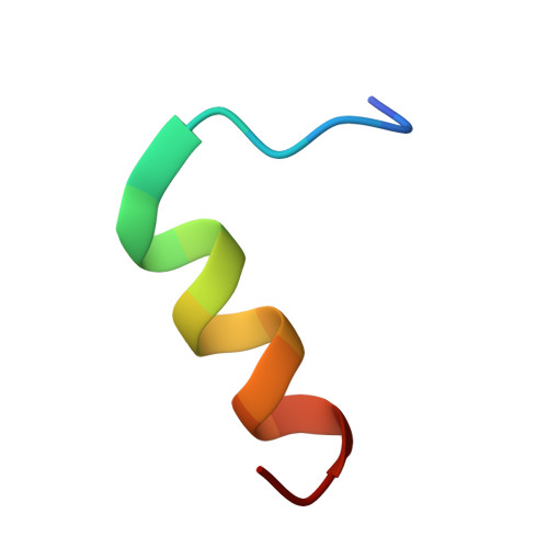
2PL9

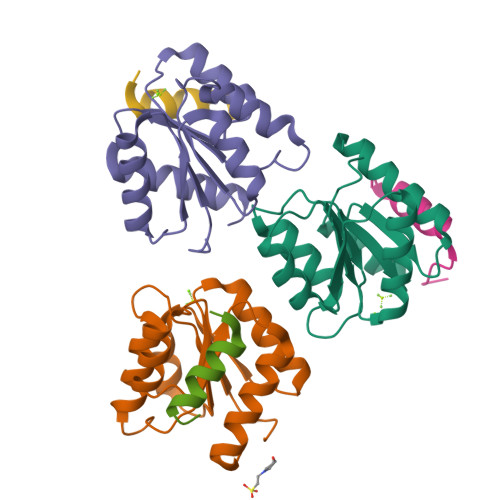
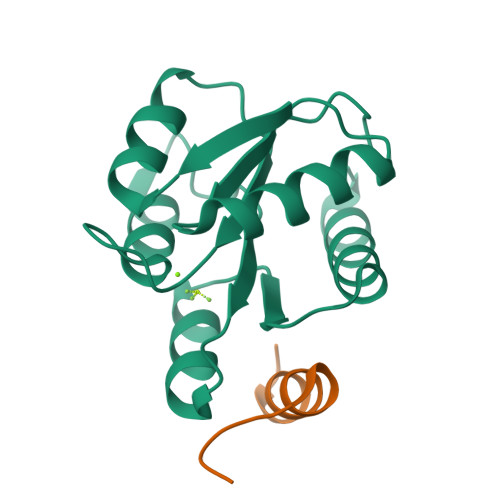
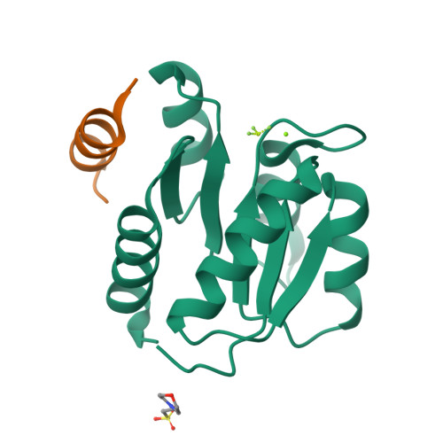
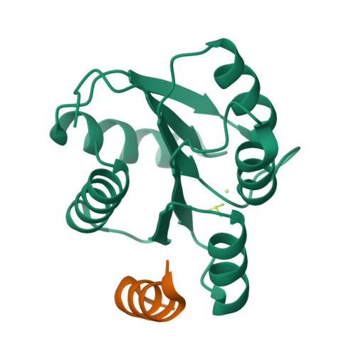
Crystal Structure of CheY-Mg(2+)-BeF(3)(-) in Complex with CheZ(C19) Peptide solved from a P2(1)2(1)2 Crystal


Experimental Data Snapshot

  • Method: X-RAY DIFFRACTION
  • Resolution: 2.60 Å
  • R-Value Free: 0.274 
  • R-Value Work: 0.213 
  • R-Value Observed: 0.220 

Starting Model: experimental
View more details

wwPDB Validation   3D Report Full Report


This is version 1.5 of the entry. See complete history


Literature

Interaction of CheY with the C-terminal peptide of CheZ.

Guhaniyogi, J.Wu, T.Patel, S.S.Stock, A.M.

(2008) J Bacteriol 190: 1419-1428

  • DOI: https://doi.org/10.1128/JB.01414-07
  • Primary Citation of Related Structures:  
    2PL9, 2PMC

  • PubMed Abstract: 

    Chemotaxis, a means for motile bacteria to sense the environment and achieve directed swimming, is controlled by flagellar rotation. The primary output of the chemotaxis machinery is the phosphorylated form of the response regulator CheY (P-CheY). The steady-state level of P-CheY dictates the direction of rotation of the flagellar motor. The chemotaxis signal in the form of P-CheY is terminated by the phosphatase CheZ. Efficient dephosphorylation of CheY by CheZ requires two distinct protein-protein interfaces: one involving the strongly conserved C-terminal helix of CheZ (CheZ(C)) tethering the two proteins together and the other constituting an active site for catalytic dephosphorylation. In a previous work (J. Guhaniyogi, V. L. Robinson, and A. M. Stock, J. Mol. Biol. 359:624-645, 2006), we presented high-resolution crystal structures of CheY in complex with the CheZ(C) peptide that revealed alternate binding modes subject to the conformational state of CheY. In this study, we report biochemical and structural data that support the alternate-binding-mode hypothesis and identify key recognition elements in the CheY-CheZ(C) interaction. In addition, we present kinetic studies of the CheZ(C)-associated effect on CheY phosphorylation with its physiologically relevant phosphodonor, the histidine kinase CheA. Our results indicate mechanistic differences in phosphotransfer from the kinase CheA versus that from small-molecule phosphodonors, explaining a modest twofold increase of CheY phosphorylation with the former, observed in this study, relative to a 10-fold increase previously documented with the latter.


  • Organizational Affiliation

    Center for Advanced Biotechnology and Medicine, 679 Hoes Lane, Piscataway, NJ 08854, USA.


Macromolecules
Find similar proteins by:  (by identity cutoff)  |  3D Structure
Entity ID: 1
MoleculeChains Sequence LengthOrganismDetailsImage
Chemotaxis protein cheY
A, B, C
128Salmonella enterica subsp. enterica serovar Typhimurium str. LT2Mutation(s): 0 
Gene Names: cheY
UniProt
Find proteins for P0A2D5 (Salmonella typhimurium (strain LT2 / SGSC1412 / ATCC 700720))
Explore P0A2D5 
Go to UniProtKB:  P0A2D5
Entity Groups  
Sequence Clusters30% Identity50% Identity70% Identity90% Identity95% Identity100% Identity
UniProt GroupP0A2D5
Sequence Annotations
Expand
  • Reference Sequence

Find similar proteins by:  Sequence   |   3D Structure  

Entity ID: 2
MoleculeChains Sequence LengthOrganismDetailsImage
Chemotaxis protein cheZ
D, E, F
19N/AMutation(s): 0 
EC: 3.1.3
UniProt
Find proteins for P07800 (Salmonella typhimurium (strain LT2 / SGSC1412 / ATCC 700720))
Explore P07800 
Go to UniProtKB:  P07800
Entity Groups  
Sequence Clusters30% Identity50% Identity70% Identity90% Identity95% Identity100% Identity
UniProt GroupP07800
Sequence Annotations
Expand
  • Reference Sequence
Small Molecules
Ligands 3 Unique
IDChains Name / Formula / InChI Key2D Diagram3D Interactions
MES
Query on MES

Download Ideal Coordinates CCD File 
K [auth B]2-(N-MORPHOLINO)-ETHANESULFONIC ACID
C6 H13 N O4 S
SXGZJKUKBWWHRA-UHFFFAOYSA-N
BEF
Query on BEF

Download Ideal Coordinates CCD File 
H [auth A],
J [auth B],
M [auth C]
BERYLLIUM TRIFLUORIDE ION
Be F3
OGIAHMCCNXDTIE-UHFFFAOYSA-K
MG
Query on MG

Download Ideal Coordinates CCD File 
G [auth A],
I [auth B],
L [auth C]
MAGNESIUM ION
Mg
JLVVSXFLKOJNIY-UHFFFAOYSA-N
Experimental Data & Validation

Experimental Data

  • Method: X-RAY DIFFRACTION
  • Resolution: 2.60 Å
  • R-Value Free: 0.274 
  • R-Value Work: 0.213 
  • R-Value Observed: 0.220 
  • Space Group: P 21 21 2
Unit Cell:
Length ( Å )Angle ( ˚ )
a = 68.058α = 90
b = 162.972β = 90
c = 37.341γ = 90
Software Package:
Software NamePurpose
DENZOdata reduction
SCALEPACKdata scaling
PHASERphasing
DMphasing
REFMACrefinement
PDB_EXTRACTdata extraction
MAR345data collection

Structure Validation

View Full Validation Report


Entry History 

Deposition Data

Revision History  (Full details and data files)

  • Version 1.0: 2008-01-15
    Type: Initial release
  • Version 1.1: 2011-07-13
    Changes: Source and taxonomy, Version format compliance
  • Version 1.2: 2013-03-06
    Changes: Advisory, Other
  • Version 1.3: 2017-10-18
    Changes: Refinement description
  • Version 1.4: 2020-09-02
    Changes: Data collection, Derived calculations, Structure summary
  • Version 1.5: 2023-08-30
    Changes: Data collection, Database references, Refinement description