2PK0

Structure of the S. agalactiae serine/threonine phosphatase at 2.65 resolution


Experimental Data Snapshot

  • Method: X-RAY DIFFRACTION
  • Resolution: 2.65 Å
  • R-Value Free: 0.271 
  • R-Value Work: 0.197 
  • R-Value Observed: 0.200 

Starting Model: experimental
View more details

wwPDB Validation   3D Report Full Report


This is version 1.4 of the entry. See complete history


Literature

Structure of Streptococcus agalactiae serine/threonine phosphatase. The subdomain conformation is coupled to the binding of a third metal ion

Rantanen, M.K.Lehtio, L.Rajagopal, L.Rubens, C.E.Goldman, A.

(2007) FEBS J 274: 3128-3137

  • DOI: https://doi.org/10.1111/j.1742-4658.2007.05845.x
  • Primary Citation of Related Structures:  
    2PK0

  • PubMed Abstract: 

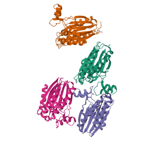
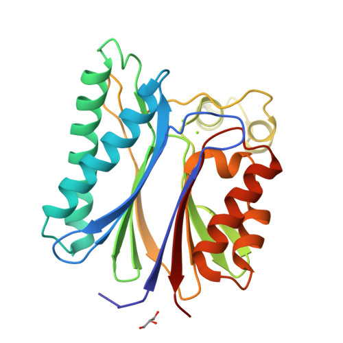
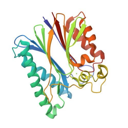
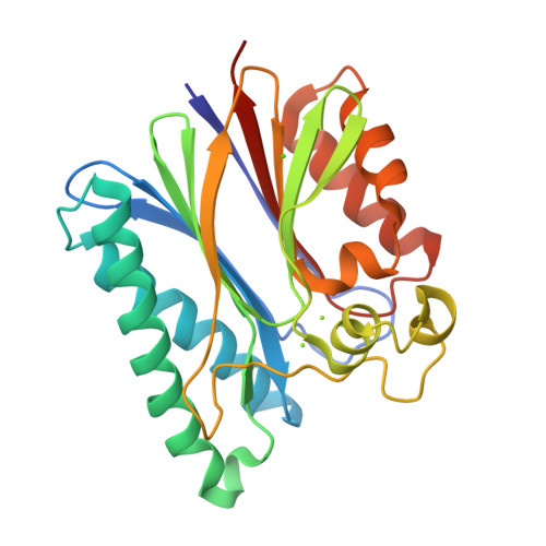
    We solved the crystal structure of Streptococcus agalactiae serine/threonine phosphatase (SaSTP) using a combination of single-wavelength anomalous dispersion phasing and molecular replacement. The overall structure resembles that of previously characterized members of the PPM/PP2C STP family. The asymmetric unit contains four monomers and we observed two novel conformations for the flap domain among them. In one of these conformations, the enzyme binds three metal ions, whereas in the other it binds only two. The three-metal ion structure also has the active site arginine in a novel conformation. The switch between the two- and three-metal ion structures appears to be binding of another monomer to the active site of STP, which promotes binding of the third metal ion. This interaction may mimic the binding of a product complex, especially since the motif binding to the active site contains a serine residue aligning remarkably well with the phosphate found in the human STP structure.


  • Organizational Affiliation

    Institute of Biotechnology, University of Helsinki, Finland, and Division of Infectious Disease, Children's Hospital and Regional Medical Center, Seattle, WA, USA.


Macromolecules
Find similar proteins by:  (by identity cutoff)  |  3D Structure
Entity ID: 1
MoleculeChains Sequence LengthOrganismDetailsImage
Serine/threonine protein phosphatase Stp1
A, B, C, D
250Streptococcus agalactiae A909Mutation(s): 0 
Gene Names: stp
EC: 3.1.3.16
UniProt
Find proteins for Q8VQA1 (Streptococcus agalactiae)
Explore Q8VQA1 
Go to UniProtKB:  Q8VQA1
Entity Groups  
Sequence Clusters30% Identity50% Identity70% Identity90% Identity95% Identity100% Identity
UniProt GroupQ8VQA1
Sequence Annotations
Expand
  • Reference Sequence
Small Molecules
Ligands 3 Unique
IDChains Name / Formula / InChI Key2D Diagram3D Interactions
GOL
Query on GOL

Download Ideal Coordinates CCD File 
I [auth B]GLYCEROL
C3 H8 O3
PEDCQBHIVMGVHV-UHFFFAOYSA-N
CL
Query on CL

Download Ideal Coordinates CCD File 
P [auth D]CHLORIDE ION
Cl
VEXZGXHMUGYJMC-UHFFFAOYSA-M
MG
Query on MG

Download Ideal Coordinates CCD File 
E [auth A]
F [auth A]
G [auth B]
H [auth B]
J [auth C]
E [auth A],
F [auth A],
G [auth B],
H [auth B],
J [auth C],
K [auth C],
L [auth C],
M [auth D],
N [auth D],
O [auth D]
MAGNESIUM ION
Mg
JLVVSXFLKOJNIY-UHFFFAOYSA-N
Experimental Data & Validation

Experimental Data

  • Method: X-RAY DIFFRACTION
  • Resolution: 2.65 Å
  • R-Value Free: 0.271 
  • R-Value Work: 0.197 
  • R-Value Observed: 0.200 
  • Space Group: P 21 21 2
Unit Cell:
Length ( Å )Angle ( ˚ )
a = 139.4α = 90
b = 92.1β = 90
c = 86.9γ = 90
Software Package:
Software NamePurpose
REFMACrefinement
MAR345dtbdata collection
XDSdata reduction
XSCALEdata scaling
MOLREPphasing

Structure Validation

View Full Validation Report


Entry History 

Deposition Data

Revision History  (Full details and data files)

  • Version 1.0: 2007-06-12
    Type: Initial release
  • Version 1.1: 2008-05-01
    Changes: Version format compliance
  • Version 1.2: 2011-07-13
    Changes: Source and taxonomy, Version format compliance
  • Version 1.3: 2024-03-13
    Changes: Data collection, Database references, Derived calculations, Refinement description
  • Version 1.4: 2024-04-03
    Changes: Refinement description