2PJV | pdb_00002pjv

solution structure of hiv-1 gp41 fusion domain bound to DPC micelle


Experimental Data Snapshot

  • Method: SOLUTION NMR
  • Conformers Calculated: 38 
  • Conformers Submitted: 38 
  • Selection Criteria: structures with the lowest energy 

wwPDB Validation 3D Report Full Report

Validation slider image for 2PJV

This is version 1.5 of the entry. See complete history

Literature

Structure and Plasticity of the Human Immunodeficiency Virus gp41 Fusion Domain in Lipid Micelles and Bilayers.

Li, Y.Tamm, L.K.

(2007) Biophys J 93: 876-885

  • DOI: https://doi.org/10.1529/biophysj.106.102335
  • Primary Citation Related Structures: 
    2PJV

  • PubMed Abstract: 

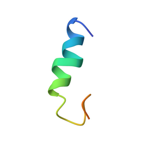
    A thorough understanding of the structure of fusion domains of enveloped viruses in changing lipid environments helps us to formulate mechanistic models on how they might function in mediating viral entry by membrane fusion. We have expressed the N-terminal fusion domain of HIV-1 gp41 as a construct that is water-soluble in the absence of membranes, but that also binds with high affinity to lipid micelles and bilayers in their presence. We have solved the structure and studied the dynamics of this domain bound to dodecylphosphocholine micelles by homo- and heteronuclear NMR spectroscopy. The fusion peptide forms a stable hydrophobic helix from Ile(4) to Ala(14), but is increasingly more disordered and dynamic in a segment of intermediate polarity that stretches from Ala(15) to Ser(23). When bound to lipid bilayers at low concentration, the HIV fusion domain is also largely alpha-helical, as determined by CD and FTIR spectroscopy. However, at higher protein/lipid ratios, the domain is partially converted to form beta-structures in lipid bilayers. Controlled lipid mixing occurs at concentrations that support the alpha-helical, but not the beta-strand conformation.


  • Organizational Affiliation
    • Department of Molecular Physiology and Biological Physics, University of Virginia, Charlottesville, Virginia, USA.

Macromolecule Content 

  • Total Structure Weight: 2.82 kDa 
  • Atom Count: 145 
  • Modeled Residue Count: 24 
  • Deposited Residue Count: 31 
  • Unique protein chains: 1

Macromolecules

Find similar proteins by:|  3D Structure
Entity ID: 1
MoleculeChains  Sequence LengthOrganismDetailsImage
Envelope glycoprotein31Human immunodeficiency virus 1Mutation(s): 1 
Gene Names: env
Membrane Entity: Yes 
UniProt
Find proteins for P19551 (Human immunodeficiency virus type 1 group M subtype B (isolate MFA))
Explore P19551 
Go to UniProtKB:  P19551
Entity Groups
Sequence Clusters30% Identity50% Identity70% Identity90% Identity95% Identity100% Identity
UniProt GroupP19551
Sequence Annotations
Expand
Reference Sequence

Experimental Data & Validation

Experimental Data

  • Method: SOLUTION NMR
  • Conformers Calculated: 38 
  • Conformers Submitted: 38 
  • Selection Criteria: structures with the lowest energy 

Structure Validation

View Full Validation Report


Entry History 

Deposition Data

Revision History  (Full details and data files)

  • Version 1.0: 2007-05-01
    Type: Initial release
  • Version 1.1: 2008-05-01
    Changes: Version format compliance
  • Version 1.2: 2011-07-13
    Changes: Version format compliance
  • Version 1.3: 2018-01-31
    Changes: Derived calculations, Structure summary
  • Version 1.4: 2021-10-20
    Changes: Data collection, Database references
  • Version 1.5: 2024-05-22
    Changes: Data collection