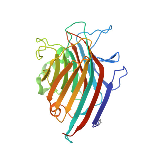
2PHU

Pterocarpus angolensis lectin in complex with Man-8D1D3


Experimental Data Snapshot

  • Method: X-RAY DIFFRACTION
  • Resolution: 2.20 Å
  • R-Value Free: 0.209 
  • R-Value Work: 0.174 

Starting Model: experimental
View more details

wwPDB Validation   3D Report Full Report


Ligand Structure Quality Assessment 


This is version 3.2 of the entry. See complete history


Literature

How a plant lectin recognizes high mannose oligosaccharides

Garcia-Pino, A.Buts, L.Wyns, L.Imberty, A.Loris, R.

(2007) Plant Physiol 144: 1733-1741

  • DOI: https://doi.org/10.1104/pp.107.100867
  • Primary Citation of Related Structures:  
    2PHF, 2PHR, 2PHT, 2PHU, 2PHW, 2PHX

  • PubMed Abstract: 

    The crystal structure of Pterocarpus angolensis seed lectin is presented in complex with a series of high mannose (Man) oligosaccharides ranging from Man-5 to Man-9. Despite that several of the nine Man residues of Man-9 have the potential to bind in the monosaccharide-binding site, all oligomannoses are bound in the same unique way, employing the tetrasaccharide sequence Manalpha(1-2)Manalpha(1-6)[Manalpha(1-3)]Manalpha(1-. Isothermal titration calorimetry titration experiments using Man-5, Man-9, and the Man-9-containing glycoprotein soybean (Glycine max) agglutinin as ligands confirm the monovalence of Man-9 and show a 4-times higher affinity for Man-9 when it is presented to P. angolensis seed lectin in a glycoprotein context.


  • Organizational Affiliation

    Laboratorium voor Ultrastructuur, Vrije Universiteit Brussel, Pleinlaan 2, B-1050 Brussel, Belgium.


Macromolecules
Find similar proteins by:  (by identity cutoff)  |  3D Structure
Entity ID: 1
MoleculeChains Sequence LengthOrganismDetailsImage
Lectin
A, B
252Pterocarpus angolensisMutation(s): 0 
UniProt
Find proteins for Q8GSD2 (Pterocarpus angolensis)
Explore Q8GSD2 
Go to UniProtKB:  Q8GSD2
Entity Groups  
Sequence Clusters30% Identity50% Identity70% Identity90% Identity95% Identity100% Identity
UniProt GroupQ8GSD2
Sequence Annotations
Expand
  • Reference Sequence
Oligosaccharides

Help

Entity ID: 2
MoleculeChains Length2D Diagram Glycosylation3D Interactions
alpha-D-mannopyranose-(1-2)-alpha-D-mannopyranose-(1-6)-[alpha-D-mannopyranose-(1-3)]alpha-D-mannopyranose-(1-6)-beta-D-mannopyranose
C
5N/A
Glycosylation Resources
GlyTouCan:  G11308TN
GlyCosmos:  G11308TN
GlyGen:  G11308TN
Small Molecules
Modified Residues  1 Unique
IDChains TypeFormula2D DiagramParent
PCA
Query on PCA
A, B
L-PEPTIDE LINKINGC5 H7 N O3GLN
Experimental Data & Validation

Experimental Data

  • Method: X-RAY DIFFRACTION
  • Resolution: 2.20 Å
  • R-Value Free: 0.209 
  • R-Value Work: 0.174 
  • Space Group: P 21 21 21
Unit Cell:
Length ( Å )Angle ( ˚ )
a = 56.696α = 90
b = 83.348β = 90
c = 122.69γ = 90
Software Package:
Software NamePurpose
MAR345data collection
CNSrefinement
DENZOdata reduction
SCALEPACKdata scaling

Structure Validation

View Full Validation Report


Ligand Structure Quality Assessment 


Entry History 

Deposition Data

Revision History  (Full details and data files)

  • Version 1.0: 2007-07-10
    Type: Initial release
  • Version 1.1: 2008-05-01
    Changes: Version format compliance
  • Version 1.2: 2011-07-13
    Changes: Version format compliance
  • Version 1.3: 2017-10-18
    Changes: Advisory, Refinement description
  • Version 2.0: 2019-12-25
    Changes: Data collection, Database references, Derived calculations, Polymer sequence
  • Version 3.0: 2020-07-29
    Type: Remediation
    Reason: Carbohydrate remediation
    Changes: Atomic model, Data collection, Derived calculations, Structure summary
  • Version 3.1: 2023-08-30
    Changes: Data collection, Database references, Derived calculations, Refinement description, Structure summary
  • Version 3.2: 2024-10-16
    Changes: Structure summary