2PHA | pdb_00002pha

Crystal structure of native, unliganded human arginase at 1.90 resolution


Experimental Data Snapshot

  • Method: X-RAY DIFFRACTION
  • Resolution: 1.90 Å
  • R-Value Free: 
    0.244 (Depositor) 
  • R-Value Work: 
    0.198 (Depositor), 0.197 (DCC) 

Starting Model: experimental
View more details

wwPDB Validation 3D Report Full Report

Validation slider image for 2PHA

This is version 1.3 of the entry. See complete history

Literature

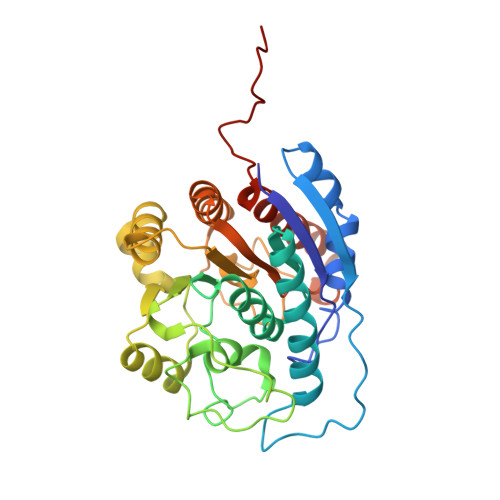
Crystal structure of human arginase I complexed with thiosemicarbazide reveals an unusual thiocarbonyl mu-sulfide ligand in the binuclear manganese cluster.

Di Costanzo, L.Pique, M.E.Christianson, D.W.

(2007) J Am Chem Soc 129: 6388-6389

  • DOI: https://doi.org/10.1021/ja071567j
  • Primary Citation Related Structures: 
    2PHA, 2PHO, 2ZAV

  • PubMed Abstract: 

    The crystal structure of the human arginase I-thiosemicarbazide complex reveals an unusual thiocarbonyl μ-sulfide ligand in the binuclear manganese cluster. The C=S moiety of thiosemicarbazide bridges Mn 2+ A and Mn 2+ B with coordination distances of 2.6 Å and 2.4 Å, respectively. Otherwise, the binding of thiosemicarbazide to human arginase I does not cause any significant structural changes in the active site. The crystal structure of the unliganded enzyme reveals a hydrogen bonded water molecule that could support proton transfer between a μ-water molecule and H141 to regenerate the nucleophilic μ-hydroxide ion in the final step of catalysis.


  • Organizational Affiliation
    • Roy and Diana Vagelos Laboratories, Department of Chemistry, University of Pennsylvania, Philadelphia, PA 19104-6323, USA.

Macromolecule Content 

  • Total Structure Weight: 69.78 kDa 
  • Atom Count: 5,032 
  • Modeled Residue Count: 628 
  • Deposited Residue Count: 644 
  • Unique protein chains: 1

Macromolecules

Find similar proteins by:|  3D Structure
Entity ID: 1
MoleculeChains  Sequence LengthOrganismDetailsImage
Arginase-1
A, B
322Homo sapiensMutation(s): 0 
Gene Names: ARG1
EC: 3.5.3.1
UniProt & NIH Common Fund Data Resources
Find proteins for P05089 (Homo sapiens)
Explore P05089 
Go to UniProtKB:  P05089
PHAROS:  P05089
GTEx:  ENSG00000118520 
Entity Groups
Sequence Clusters30% Identity50% Identity70% Identity90% Identity95% Identity100% Identity
UniProt GroupP05089
Sequence Annotations
Expand
Reference Sequence

Experimental Data & Validation

Experimental Data

  • Method: X-RAY DIFFRACTION
  • Resolution: 1.90 Å
  • R-Value Free:  0.244 (Depositor) 
  • R-Value Work:  0.198 (Depositor), 0.197 (DCC) 
Space Group: P 3
Unit Cell:
Length ( Å )Angle ( ˚ )
a = 90.77α = 90
b = 90.77β = 90
c = 69.74γ = 120
Software Package:
Software NamePurpose
ADSCdata collection
PHASERphasing
CNSrefinement
MOSFLMdata reduction
CCP4data scaling

Structure Validation

View Full Validation Report


Entry History 

Deposition Data

Revision History  (Full details and data files)

  • Version 1.0: 2007-05-08
    Type: Initial release
  • Version 1.1: 2007-12-17
    Changes: Version format compliance
  • Version 1.2: 2011-07-13
    Changes: Derived calculations, Version format compliance
  • Version 1.3: 2023-08-30
    Changes: Data collection, Database references, Derived calculations, Refinement description