2PEC | pdb_00002pec

THE REFINED THREE-DIMENSIONAL STRUCTURE OF PECTATE LYASE C FROM ERWINIA CHRYSANTHEMI AT 2.2 ANGSTROMS RESOLUTION: IMPLICATIONS FOR AN ENZYMATIC MECHANISM


Experimental Data Snapshot

  • Method: X-RAY DIFFRACTION
  • Resolution: 2.20 Å
  • R-Value Work: 
    0.180 (Depositor) 
  • R-Value Observed: 
    0.180 (Depositor) 

wwPDB Validation 3D Report Full Report

Validation slider image for 2PEC

This is version 1.4 of the entry. See complete history

Literature

Protein motifs. 3. The parallel beta helix and other coiled folds.

Yoder, M.D.Jurnak, F.

(1995) FASEB J 9: 335-342

  • DOI: https://doi.org/10.1096/fasebj.9.5.7896002
  • Primary Citation Related Structures: 
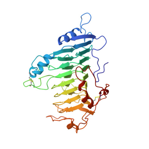
    2PEC

  • PubMed Abstract: 

    A new type of structural domain, composed of all parallel beta strands, has been observed within the last year. An analysis of the basic types suggests that there are two distinct classes: the parallel beta helices, which belong to a tri beta-strand category, and the beta roll, which belongs to a di beta-strand category. The novel structural features of each class are described and the proteins belonging to each category are summarized. Proteins with the parallel beta helix fold include three pectate lyases and the tailspike protein from P22 phage. Proteins with the beta roll fold include two alkaline proteases. Although the parallel beta composition is emphasized, the same set of proteins share another common structural feature with several other proteins containing alpha helices: the polypeptide backbone is folded into a coiled structure in which each coil has the same 3-dimensional arrangement of a group of secondary structural elements. In addition to parallel beta domains, the other groups include the alpha/beta coiled fold, as represented by ribonuclease inhibitor, and the alpha/alpha coiled fold, as represented by lipovitellin and soluble lytic transglycoslyase. Novel features of the alpha/beta and alpha/alpha coiled folds are summarized.


  • Organizational Affiliation
    • School of Biological Sciences, Division of Cell Biology and Biophysics, University of Missouri, Kansas City 64110.

Macromolecule Content 

  • Total Structure Weight: 37.73 kDa 
  • Atom Count: 2,762 
  • Modeled Residue Count: 352 
  • Deposited Residue Count: 353 
  • Unique protein chains: 1

Macromolecules

Find similar proteins by:|  3D Structure
Entity ID: 1
MoleculeChains  Sequence LengthOrganismDetailsImage
PECTATE LYASE C353Dickeya chrysanthemiMutation(s): 0 
Gene Names: PPEL410 OF PELC
EC: 4.2.2.2
UniProt
Find proteins for P11073 (Dickeya chrysanthemi)
Explore P11073 
Go to UniProtKB:  P11073
Entity Groups
Sequence Clusters30% Identity50% Identity70% Identity90% Identity95% Identity100% Identity
UniProt GroupP11073
Sequence Annotations
Expand
Reference Sequence

Experimental Data & Validation

Experimental Data

  • Method: X-RAY DIFFRACTION
  • Resolution: 2.20 Å
  • R-Value Work:  0.180 (Depositor) 
  • R-Value Observed: 0.180 (Depositor) 
Space Group: P 21 21 21
Unit Cell:
Length ( Å )Angle ( ˚ )
a = 73.38α = 90
b = 80.26β = 90
c = 95.12γ = 90
Software Package:
Software NamePurpose
X-PLORmodel building
X-PLORrefinement
X-PLORphasing

Structure Validation

View Full Validation Report


Entry History 

Deposition Data

Revision History  (Full details and data files)

  • Version 1.0: 1995-02-14
    Type: Initial release
  • Version 1.1: 2008-03-24
    Changes: Version format compliance
  • Version 1.2: 2011-07-13
    Changes: Version format compliance
  • Version 1.3: 2017-11-29
    Changes: Derived calculations, Other
  • Version 1.4: 2024-10-16
    Changes: Data collection, Database references, Structure summary