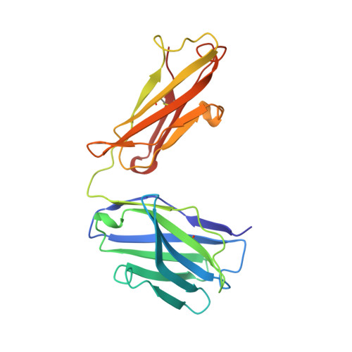
2PCP

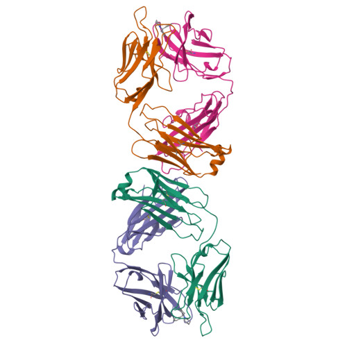
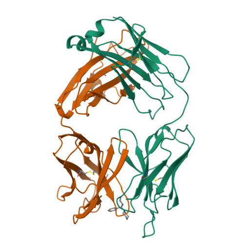
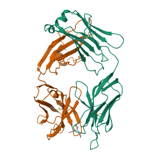
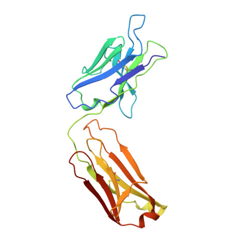
ANTIBODY FAB COMPLEXED WITH PHENCYCLIDINE


Experimental Data Snapshot

  • Method: X-RAY DIFFRACTION
  • Resolution: 2.20 Å
  • R-Value Free: 0.290 
  • R-Value Work: 0.196 
  • R-Value Observed: 0.196 

Starting Models: experimental
View more details

wwPDB Validation   3D Report Full Report


Ligand Structure Quality Assessment 


This is version 1.3 of the entry. See complete history


Literature

Crystal structure of monoclonal 6B5 Fab complexed with phencyclidine.

Lim, K.Owens, S.M.Arnold, L.Sacchettini, J.C.Linthicum, D.S.

(1998) J Biol Chem 273: 28576-28582

  • DOI: https://doi.org/10.1074/jbc.273.44.28576
  • Primary Citation of Related Structures:  
    2PCP

  • PubMed Abstract: 

    The crystal structure of monoclonal antibody (mAb) 6B5 Fab fragment complexed with 1-(1-phenylcyclohexyl)piperidine (PCP or phencyclidine) was determined at 2.2-A resolution. 6B5 was originally produced from a mouse immunized with a phencyclidine analogue hapten 5-[N-(1'phenylcyclohexyl)amino]pentanoic acid conjugated to bovine serum albumin. This mAb was selected for further study because of its high affinity (Kd = 2 x 10(-9) M/liter) for PCP and usefulness in reversing PCP-induced central nervous system toxicity in laboratory animals. The dominant feature of the 6B5 Fab.PCP complex is the deep binding site and hydrophobic nature of the interaction. The ligand binding pocket of 6B5 Fab has numerous aromatic side chains, as compared with other known Fab structures. The most notable feature of the binding site is a Trp at position 97H (H-chain), and the side chain of this residue appears to act as a hydrophobic umbrella on the ligand in the antigen binding pocket. There are only two other known Fabs found with a Trp at the 97H position in complementarity determining region (CDR) H3, but they do not play a major role in the interaction with their respective antigens; in both Fab TE33 and R6.5 the Trp 97H side chain is positioned away from the bound antigen. Comparison of the CDR residues of 6B5 with other Fab structures with similar CDR sizes and amino acid compositions reveals a number of important patterns of residue substitutions that appear to be critical for specific PCP ligand interactions.


  • Organizational Affiliation

    Center for Structural Biology, Department of Biochemistry and Biophysics, Texas A&M University, College Station, Texas, 77843, USA.


Macromolecules
Find similar proteins by:  (by identity cutoff)  |  3D Structure
Entity ID: 1
MoleculeChains Sequence LengthOrganismDetailsImage
IMMUNOGLOBULIN
A, C
216Mus musculusMutation(s): 0 
Entity Groups  
Sequence Clusters30% Identity50% Identity70% Identity90% Identity95% Identity100% Identity
Sequence Annotations
Expand
  • Reference Sequence
Find similar proteins by:  (by identity cutoff)  |  3D Structure
Entity ID: 2
MoleculeChains Sequence LengthOrganismDetailsImage
IMMUNOGLOBULIN
B, D
215Mus musculusMutation(s): 0 
Entity Groups  
Sequence Clusters30% Identity50% Identity70% Identity90% Identity95% Identity100% Identity
Sequence Annotations
Expand
  • Reference Sequence
Small Molecules
Ligands 1 Unique
IDChains Name / Formula / InChI Key2D Diagram3D Interactions
1PC
Query on 1PC

Download Ideal Coordinates CCD File 
E [auth B],
F [auth C]
1-(PHENYL-1-CYCLOHEXYL)PIPERIDINE
C17 H25 N
JTJMJGYZQZDUJJ-UHFFFAOYSA-N
Binding Affinity Annotations 
IDSourceBinding Affinity
1PC PDBBind:  2PCP Kd: 2 (nM) from 1 assay(s)
Experimental Data & Validation

Experimental Data

  • Method: X-RAY DIFFRACTION
  • Resolution: 2.20 Å
  • R-Value Free: 0.290 
  • R-Value Work: 0.196 
  • R-Value Observed: 0.196 
  • Space Group: P 1
Unit Cell:
Length ( Å )Angle ( ˚ )
a = 53.46α = 62.11
b = 67.72β = 84.66
c = 68.04γ = 83.17
Software Package:
Software NamePurpose
DENZOdata reduction
SCALEPACKdata scaling
X-PLORmodel building
X-PLORrefinement
X-PLORphasing

Structure Validation

View Full Validation Report


Ligand Structure Quality Assessment 


Entry History 

Deposition Data

Revision History  (Full details and data files)

  • Version 1.0: 1999-01-27
    Type: Initial release
  • Version 1.1: 2008-03-03
    Changes: Version format compliance
  • Version 1.2: 2011-07-13
    Changes: Version format compliance
  • Version 1.3: 2023-08-09
    Changes: Database references, Derived calculations, Refinement description