2PBJ | pdb_00002pbj

GSH-heme bound microsomal prostaglandin E synthase


Experimental Data Snapshot

  • Method: X-RAY DIFFRACTION
  • Resolution: 2.80 Å
  • R-Value Free: 
    0.269 (Depositor), 0.270 (DCC) 
  • R-Value Work: 
    0.242 (Depositor), 0.240 (DCC) 
  • R-Value Observed: 
    0.242 (Depositor) 

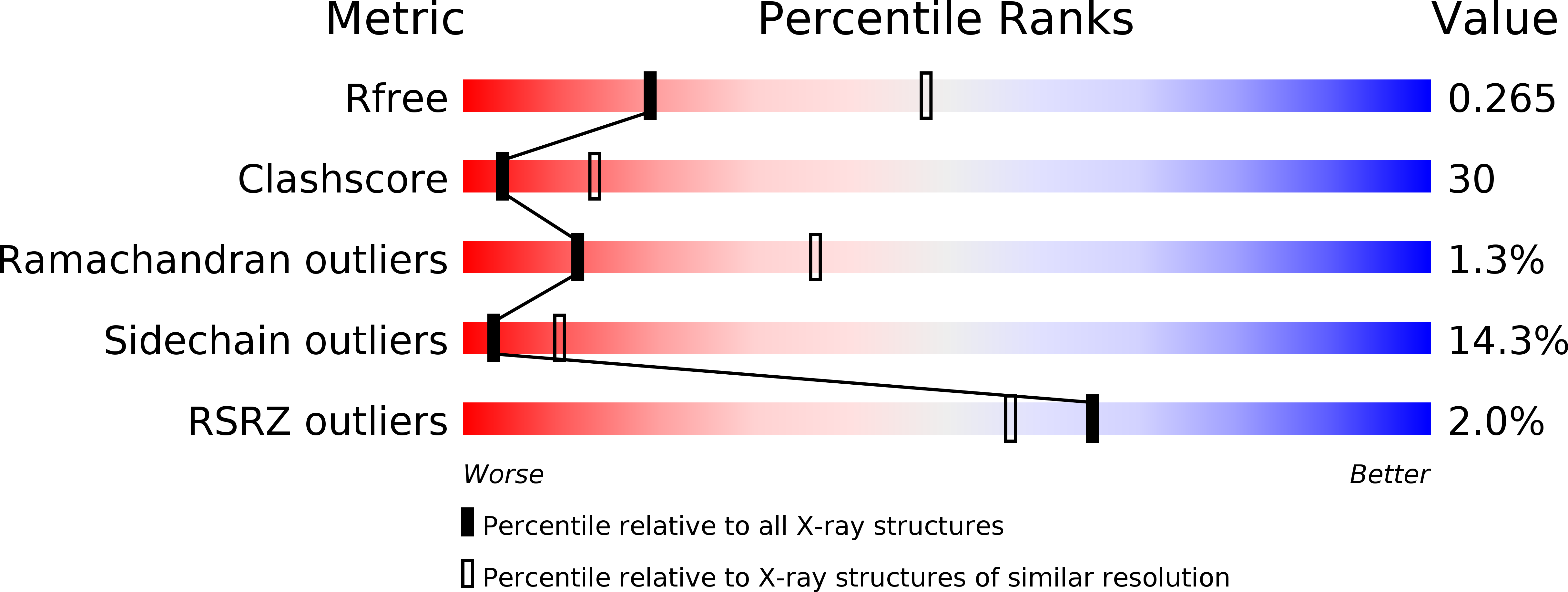
wwPDB Validation   3D Report Full Report


Ligand Structure Quality Assessment 

Created with Raphaël 2.3.0Worse 01 BetterLigand structure goodness of fit to experimental dataBest fitted HEMClick on this verticalbar to view detailsBest fitted GSHClick on this verticalbar to view details

This is version 1.2 of the entry. See complete history


Literature

PGH2 degradation pathway catalyzed by GSH-heme complex bound microsomal prostaglandin E2 synthase type 2: the first example of a dual-function enzyme.

Yamada, T.Takusagawa, F.

(2007) Biochemistry 46: 8414-8424

  • DOI: https://doi.org/10.1021/bi700605m
  • Primary Citation of Related Structures:  
    2PBJ

  • PubMed Abstract: 

    Prostaglandin E2 synthase (PGES) catalyzes the isomerization of PGH2 to PGE2. PGES type 2 (mPGES-2) is a membrane-associated enzyme, whose N-terminal section is apparently inserted into the lipid bilayer. Both intact and N-terminal truncated enzymes have been isolated and have similar catalytic activity. The recombinant N-terminal truncated enzyme purified from Escherichia coli HB101 grown in LB medium containing delta-aminolevulinate and Fe(NO3)3 has a red color, while the same enzyme purified from the same E. coli grown in minimal medium has no color. The red-colored enzyme has been characterized by mass, fluorescence, and EPR spectroscopies and X-ray crystallography. The enzyme is found to contain bound glutathione (GSH) and heme. GSH binds to the active site with six H-bonds, while a heme is complexed with bound GSH forming a S-Fe coordination bond with no polar interaction with mPGES-2. There is a large open space between the heme and the protein, where a PGH2 might be able to bind. The heme dissociation constant is 0.53 microM, indicating that mPGES-2 has relatively strong heme affinity. Indeed, expression of mPGES-2 in E. coli stimulates heme biosynthesis. Although mPGES-2 has been reported to be a GSH-independent PGES, the crystal structure and sequence analysis indicate that mPGES-2 is a GSH-binding protein. The GSH-heme complex-bound enzyme (mPGES-2h) catalyzes formation of 12(S)-hydroxy-5(Z),8(E),10(E)-heptadecatrienoic acid and malondialdehyde from PGH2, but not formation of PGE2. The following kinetic parameters at 37 degrees C were determined: KM = 56 microM, kcat = 63 s-1, and kcat/KM = 1.1 x 10(6) M-1 s-1. They suggest that mPGES-2h has significant catalytic activity for PGH2 degradation. It is possible that both GSH-heme complex-free and -bound enzymes are present in the same tissues. mPGES-2 in heme-rich liver is most likely to become the form of mPGES-2h and might be involved in degradation reactions similar to that of cytochrome P450. Since mPGES-2 is an isomerase and mPGES-2h is a lyase, mPGES-2 cannot simply be classified into one of six classes set by the International Union of Biochemistry and Molecular Biology.


  • Organizational Affiliation

    Department of Molecular Biosciences, University of Kansas, 1200 Sunnyside Avenue, Lawrence, Kansas 66045-7534, USA.


Macromolecules
Find similar proteins by:  (by identity cutoff)  |  3D Structure
Entity ID: 1
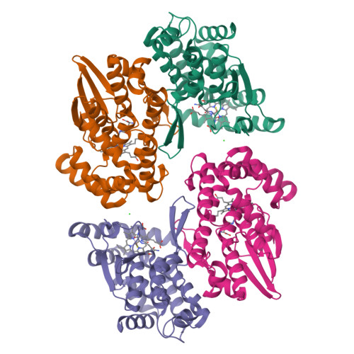
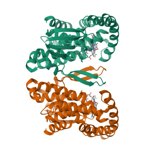
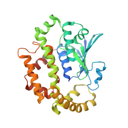
MoleculeChains Sequence LengthOrganismDetailsImage
Prostaglandin E synthase 2
A, B, C, D
290Macaca fascicularisMutation(s): 0 
Gene Names: mPGES-2
EC: 5.3.99.3
UniProt
Find proteins for Q9N0A4 (Macaca fascicularis)
Explore Q9N0A4 
Go to UniProtKB:  Q9N0A4
Entity Groups  
Sequence Clusters30% Identity50% Identity70% Identity90% Identity95% Identity100% Identity
UniProt GroupQ9N0A4
Sequence Annotations
Expand
  • Reference Sequence
Small Molecules
Ligands 3 Unique
IDChains Name / Formula / InChI Key2D Diagram3D Interactions
HEM
Query on HEM

Download Ideal Coordinates CCD File 
F [auth A],
I [auth B],
L [auth C],
O [auth D]
PROTOPORPHYRIN IX CONTAINING FE
C34 H32 Fe N4 O4
KABFMIBPWCXCRK-RGGAHWMASA-L
GSH
Query on GSH

Download Ideal Coordinates CCD File 
G [auth A],
J [auth B],
M [auth C],
P [auth D]
GLUTATHIONE
C10 H17 N3 O6 S
RWSXRVCMGQZWBV-WDSKDSINSA-N
CL
Query on CL

Download Ideal Coordinates CCD File 
E [auth A],
H [auth B],
K [auth C],
N [auth D]
CHLORIDE ION
Cl
VEXZGXHMUGYJMC-UHFFFAOYSA-M
Experimental Data & Validation

Experimental Data

  • Method: X-RAY DIFFRACTION
  • Resolution: 2.80 Å
  • R-Value Free:  0.269 (Depositor), 0.270 (DCC) 
  • R-Value Work:  0.242 (Depositor), 0.240 (DCC) 
  • R-Value Observed: 0.242 (Depositor) 
Space Group: C 1 2 1
Unit Cell:
Length ( Å )Angle ( ˚ )
a = 127α = 90
b = 122.49β = 111
c = 110.97γ = 90
Software Package:
Software NamePurpose
HKL-2000data collection
CNSrefinement
DENZOdata reduction
SCALEPACKdata scaling
CNSphasing

Structure Validation

View Full Validation Report


Ligand Structure Quality Assessment 

Created with Raphaël 2.3.0Worse 01 BetterLigand structure goodness of fit to experimental dataBest fitted HEMClick on this verticalbar to view detailsBest fitted GSHClick on this verticalbar to view details

Entry History 

Deposition Data

Revision History  (Full details and data files)

  • Version 1.0: 2008-02-12
    Type: Initial release
  • Version 1.1: 2011-07-13
    Changes: Version format compliance
  • Version 1.2: 2024-02-21
    Changes: Data collection, Database references, Derived calculations