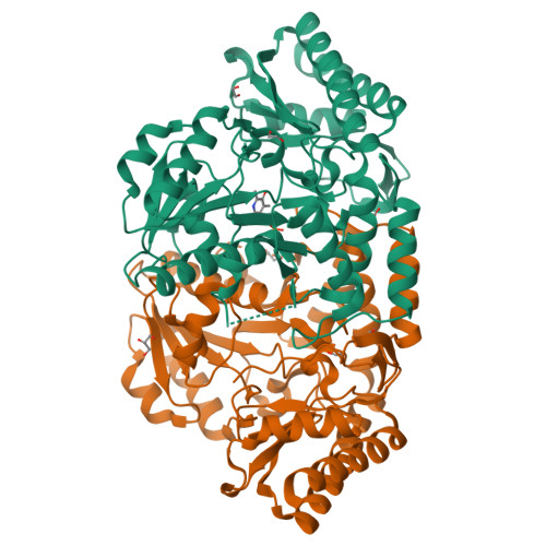
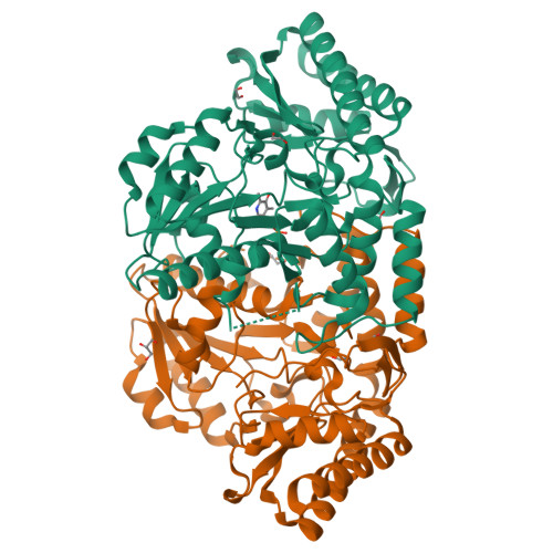
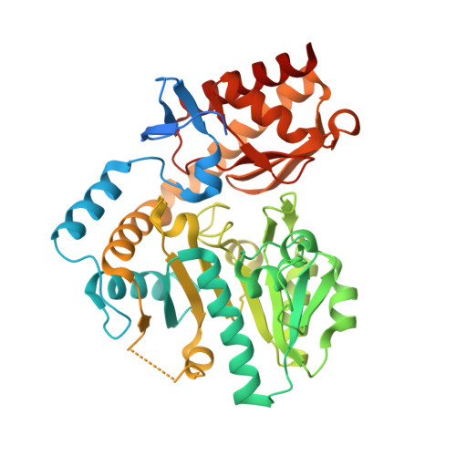
2PB2

Structure of biosynthetic N-acetylornithine aminotransferase from Salmonella typhimurium: studies on substrate specificity and inhibitor binding


Experimental Data Snapshot

  • Method: X-RAY DIFFRACTION
  • Resolution: 1.91 Å
  • R-Value Free: 0.237 
  • R-Value Work: 0.196 
  • R-Value Observed: 0.198 

Starting Model: experimental
View more details

wwPDB Validation   3D Report Full Report


Ligand Structure Quality Assessment 


This is version 1.3 of the entry. See complete history


Literature

Structure of biosynthetic N-acetylornithine aminotransferase from Salmonella typhimurium: Studies on substrate specificity and inhibitor binding

Rajaram, V.Ratna Prasuna, P.Savithri, H.S.Murthy, M.R.N.

(2007) Proteins 70: 429-441

  • DOI: https://doi.org/10.1002/prot.21567
  • Primary Citation of Related Structures:  
    2PB0, 2PB2

  • PubMed Abstract: 

    Acetylornithine aminotransferase (AcOAT) is one of the key enzymes involved in arginine metabolism and catalyzes the conversion of N-acetylglutamate semialdehyde to N-acetylornithine (AcOrn) in the presence of L-glutamate. It belongs to the Type I subgroup II family of pyridoxal 5'-phosphate (PLP) dependent enzymes. E. coli biosynthetic AcOAT (eAcOAT) also catalyzes the conversion of N-succinyl-L-2-amino-6-oxopimelate to N-succinyl-L,L-diaminopimelate, one of the steps in lysine biosynthesis. In view of the critical role of AcOAT in lysine and arginine biosynthesis, structural studies were initiated on the enzyme from S. typhimurium (sAcOAT). The K(m) and k(cat)/K(m) values determined with the purified sAcOAT suggested that the enzyme had much higher affinity for AcOrn than for ornithine (Orn) and was more efficient than eAcOAT. sAcOAT was inhibited by gabaculine (Gcn) with an inhibition constant (K(i)) of 7 microM and a second-order rate constant (k(2)) of 0.16 mM(-1) s(-1). sAcOAT, crystallized in the unliganded form and in the presence of Gcn or L-glutamate, diffracted to a maximum resolution of 1.90 A and contained a dimer in the asymmetric unit. The structure of unliganded sAcOAT showed significant electron density for PLP in only one of the subunits (subunit A). The asymmetry in PLP binding could be attributed to the ordering of the loop L(alphak-) (betam) in only one subunit (subunit B; the loop from subunit B comes close to the phosphate group of PLP in subunit A). Structural and spectral studies of sAcOAT with Gcn suggested that the enzyme might have a low affinity for PLP-Gcn complex. Comparison of sAcOAT with T. thermophilus AcOAT and human ornithine aminotransferase suggested that the higher specificity of sAcOAT towards AcOrn may not be due to specific changes in the active site residues but could result from minor conformational changes in some of them. This is the first structural report of AcOAT from a mesophilic organism and could serve as a basis for drug design as the enzyme is important for bacterial cell wall biosynthesis.


  • Organizational Affiliation

    Molecular Biophysics Unit, Indian Institute of Science, Bangalore 560012, India.


Macromolecules
Find similar proteins by:  (by identity cutoff)  |  3D Structure
Entity ID: 1
MoleculeChains Sequence LengthOrganismDetailsImage
Acetylornithine/succinyldiaminopimelate aminotransferase
A, B
420Salmonella enterica subsp. enterica serovar Typhimurium str. LT2Mutation(s): 0 
Gene Names: argD
EC: 2.6.1.11 (PDB Primary Data), 2.6.1.17 (PDB Primary Data)
UniProt
Find proteins for P40732 (Salmonella typhimurium (strain LT2 / SGSC1412 / ATCC 700720))
Explore P40732 
Go to UniProtKB:  P40732
Entity Groups  
Sequence Clusters30% Identity50% Identity70% Identity90% Identity95% Identity100% Identity
UniProt GroupP40732
Sequence Annotations
Expand
  • Reference Sequence
Small Molecules
Ligands 3 Unique
IDChains Name / Formula / InChI Key2D Diagram3D Interactions
PLP
Query on PLP

Download Ideal Coordinates CCD File 
C [auth A]PYRIDOXAL-5'-PHOSPHATE
C8 H10 N O6 P
NGVDGCNFYWLIFO-UHFFFAOYSA-N
GOL
Query on GOL

Download Ideal Coordinates CCD File 
J [auth B]GLYCEROL
C3 H8 O3
PEDCQBHIVMGVHV-UHFFFAOYSA-N
EDO
Query on EDO

Download Ideal Coordinates CCD File 
D [auth A]
E [auth A]
F [auth A]
G [auth A]
H [auth B]
D [auth A],
E [auth A],
F [auth A],
G [auth A],
H [auth B],
I [auth B]
1,2-ETHANEDIOL
C2 H6 O2
LYCAIKOWRPUZTN-UHFFFAOYSA-N
Experimental Data & Validation

Experimental Data

  • Method: X-RAY DIFFRACTION
  • Resolution: 1.91 Å
  • R-Value Free: 0.237 
  • R-Value Work: 0.196 
  • R-Value Observed: 0.198 
  • Space Group: P 21 21 2
Unit Cell:
Length ( Å )Angle ( ˚ )
a = 97.065α = 90
b = 111.966β = 90
c = 65.489γ = 90
Software Package:
Software NamePurpose
REFMACrefinement
MAR345dtbdata collection
DENZOdata reduction
SCALEPACKdata scaling
AMoREphasing

Structure Validation

View Full Validation Report


Ligand Structure Quality Assessment 


Entry History 

Deposition Data

Revision History  (Full details and data files)

  • Version 1.0: 2007-12-25
    Type: Initial release
  • Version 1.1: 2011-07-13
    Changes: Source and taxonomy, Version format compliance
  • Version 1.2: 2024-03-13
    Changes: Data collection, Database references, Derived calculations
  • Version 1.3: 2024-04-03
    Changes: Refinement description