2PA2 | pdb_00002pa2

Crystal structure of human Ribosomal protein L10 core domain


Experimental Data Snapshot

  • Method: X-RAY DIFFRACTION
  • Resolution: 2.50 Å
  • R-Value Free: 
    0.264 (Depositor) 
  • R-Value Work: 
    0.240 (Depositor), 0.241 (DCC) 
  • R-Value Observed: 
    0.240 (Depositor) 

Starting Model: experimental
View more details

wwPDB Validation 3D Report Full Report

Validation slider image for 2PA2

This is version 1.3 of the entry. See complete history

Literature

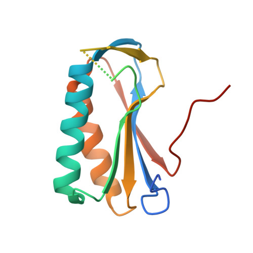
Crystal Structure of Human Ribosomal Protein L10 Core Domain Reveals Eukaryote-Specific Motifs in Addition to the Conserved Fold

Nishimura, M.Kaminishi, T.Takemoto, C.Kawazoe, M.Yoshida, T.Tanaka, A.Sugano, S.Shirouzu, M.Ohkubo, T.Yokoyama, S.Kobayashi, Y.

(2008) J Mol Biology 377: 421-430

  • DOI: https://doi.org/10.1016/j.jmb.2008.01.003
  • Primary Citation Related Structures: 
    2PA2

  • PubMed Abstract: 

    A phylogenetically conserved ribosomal protein L16p/L10e organizes the architecture of the aminoacyl tRNA binding site on the large ribosomal subunit. Eukaryotic L10 also exhibits a variety of cellular activities, and, in particular, human L10 is known as a putative tumor suppressor, QM. We have determined the 2.5-A crystal structure of the human L10 core domain that corresponds to residues 34-182 of the full-length 214 amino acids. Its two-layered alpha+beta architecture is significantly similar to those of the archaeal and bacterial homologues, substantiating a high degree of structural conservation across the three phylogenetic domains. A cation-binding pocket formed between alpha2 and beta 6 is similar to that of the archaeal L10 protein but appears to be better ordered. Previously reported L10 mutations that cause defects in the yeast ribosome are clustered around this pocket, indicating that its integrity is crucial for its role in L10 function. Characteristic interactions among Arg90-Trp171-Arg139 guide the C-terminal part outside of the central fold, implying that the eukaryote-specific C-terminal extension localizes on the outer side of the ribosome.


  • Organizational Affiliation
    • Graduate School of Pharmaceutical Sciences, Osaka University, 1-6 Yamadaoka, Suita, Osaka 565-0871, Japan.

Macromolecule Content 

  • Total Structure Weight: 34.47 kDa 
  • Atom Count: 1,787 
  • Modeled Residue Count: 216 
  • Deposited Residue Count: 302 
  • Unique protein chains: 1

Macromolecules

Find similar proteins by:|  3D Structure
Entity ID: 1
MoleculeChains  Sequence LengthOrganismDetailsImage
60S ribosomal protein L10
A, B
151Homo sapiensMutation(s): 0 
UniProt & NIH Common Fund Data Resources
Find proteins for P27635 (Homo sapiens)
Explore P27635 
Go to UniProtKB:  P27635
PHAROS:  P27635
GTEx:  ENSG00000147403 
Entity Groups
Sequence Clusters30% Identity50% Identity70% Identity90% Identity95% Identity100% Identity
UniProt GroupP27635
Sequence Annotations
Expand
Reference Sequence

Experimental Data & Validation

Experimental Data

  • Method: X-RAY DIFFRACTION
  • Resolution: 2.50 Å
  • R-Value Free:  0.264 (Depositor) 
  • R-Value Work:  0.240 (Depositor), 0.241 (DCC) 
  • R-Value Observed: 0.240 (Depositor) 
Space Group: P 31 2 1
Unit Cell:
Length ( Å )Angle ( ˚ )
a = 52.724α = 90
b = 52.724β = 90
c = 185.489γ = 120
Software Package:
Software NamePurpose
CNSrefinement
ADSCdata collection
HKL-2000data reduction
HKL-2000data scaling
PHASERphasing

Structure Validation

View Full Validation Report


Entry History 

Revision History  (Full details and data files)

  • Version 1.0: 2008-03-11
    Type: Initial release
  • Version 1.1: 2011-07-13
    Changes: Version format compliance
  • Version 1.2: 2019-11-06
    Changes: Data collection, Database references, Structure summary
  • Version 1.3: 2023-10-25
    Changes: Data collection, Database references, Derived calculations, Refinement description