Rational design of a novel, potent, and orally bioavailable cyclohexylamine DPP-4 inhibitor by application of molecular modeling and X-ray crystallography of sitagliptin
Biftu, T., Scapin, G., Singh, S., Feng, D., Becker, J.W., Eiermann, G., He, H., Lyons, K., Patel, S., Petrov, A., Sinha-Roy, R., Zhang, B., Wu, J., Zhang, X., Doss, G.A., Thornberry, N.A., Weber, A.E.(2007) Bioorg Med Chem Lett 17: 3384-3387
- PubMed: 17433672 Search on PubMed
- DOI: https://doi.org/10.1016/j.bmcl.2007.03.095
- Primary Citation Related Structures:
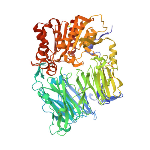
2P8S - PubMed Abstract:
Molecular modeling was used to design a rigid analog of sitagliptin 1. The X-ray crystal structure of sitagliptin bound to DPP-4 suggested that the central beta-amino butyl amide moiety could be replaced with a cyclohexylamine group. This was confirmed by structural analysis and the resulting analog 2a was synthesized and found to be a potent DPP-4 inhibitor (IC(50)=21 nM) with excellent in vivo activity and pharmacokinetic profile.
- Department of Medicinal Chemistry, Merck Research Laboratories, Merck & Co., Inc., PO Box 2000, Rahway, NJ 07065, USA. Tesfaye_Biftu@merck.com
Organizational Affiliation: