2P7U | pdb_00002p7u

The crystal structure of rhodesain, the major cysteine protease of T. brucei rhodesiense, bound to inhibitor K777


Experimental Data Snapshot

  • Method: X-RAY DIFFRACTION
  • Resolution: 1.65 Å
  • R-Value Free: 
    0.175 (Depositor), 0.182 (DCC) 
  • R-Value Work: 
    0.135 (Depositor), 0.145 (DCC) 
  • R-Value Observed: 
    0.137 (Depositor) 

Starting Model: experimental
View more details

wwPDB Validation 3D Report Full Report

Validation slider image for 2P7U

Ligand Structure Quality Assessment 


This is version 1.4 of the entry. See complete history

Literature

Vinyl sulfones as antiparasitic agents and a structural basis for drug design.

Kerr, I.D.Lee, J.H.Farady, C.J.Marion, R.Rickert, M.Sajid, M.Pandey, K.C.Caffrey, C.R.Legac, J.Hansell, E.McKerrow, J.H.Craik, C.S.Rosenthal, P.J.Brinen, L.S.

(2009) J Biological Chem 284: 25697-25703

  • DOI: https://doi.org/10.1074/jbc.M109.014340
  • Primary Citation Related Structures: 
    2OZ2, 2P7U, 3BWK

  • PubMed Abstract: 

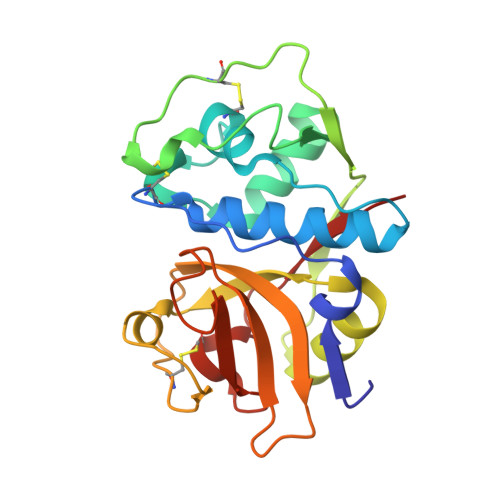
    Cysteine proteases of the papain superfamily are implicated in a number of cellular processes and are important virulence factors in the pathogenesis of parasitic disease. These enzymes have therefore emerged as promising targets for antiparasitic drugs. We report the crystal structures of three major parasite cysteine proteases, cruzain, falcipain-3, and the first reported structure of rhodesain, in complex with a class of potent, small molecule, cysteine protease inhibitors, the vinyl sulfones. These data, in conjunction with comparative inhibition kinetics, provide insight into the molecular mechanisms that drive cysteine protease inhibition by vinyl sulfones, the binding specificity of these important proteases and the potential of vinyl sulfones as antiparasitic drugs.


  • Organizational Affiliation
    • Department of Cellular and Molecular Pharmacology, University of California, San Francisco, California 94158-2550, USA.

Macromolecule Content 

  • Total Structure Weight: 23.48 kDa 
  • Atom Count: 1,836 
  • Modeled Residue Count: 215 
  • Deposited Residue Count: 215 
  • Unique protein chains: 1

Macromolecules

Find similar proteins by:|  3D Structure
Entity ID: 1
MoleculeChains  Sequence LengthOrganismDetailsImage
Cysteine protease215Trypanosoma brucei rhodesienseMutation(s): 1 
Gene Names: rhodesain
UniProt
Find proteins for Q95PM0 (Trypanosoma brucei rhodesiense)
Explore Q95PM0 
Go to UniProtKB:  Q95PM0
Entity Groups
Sequence Clusters30% Identity50% Identity70% Identity90% Identity95% Identity100% Identity
UniProt GroupQ95PM0
Sequence Annotations
Expand
Reference Sequence

Small Molecules

Ligands 1 Unique
IDChains Name / Formula / InChI Key2D Diagram3D Interactions
D1R

Query on D1R


Download:Ideal Coordinates CCD File
B [auth A]NALPHA-[(4-METHYLPIPERAZIN-1-YL)CARBONYL]-N-{(1S)-3-PHENYL-1-[2-(PHENYLSULFONYL)ETHYL]PROPYL}-L-PHENYLALANINAMIDE
C32 H40 N4 O4 S
VZSXPUDQSLKVIR-JDXGNMNLSA-N

Experimental Data & Validation

Experimental Data

  • Method: X-RAY DIFFRACTION
  • Resolution: 1.65 Å
  • R-Value Free:  0.175 (Depositor), 0.182 (DCC) 
  • R-Value Work:  0.135 (Depositor), 0.145 (DCC) 
  • R-Value Observed: 0.137 (Depositor) 
Space Group: P 1
Unit Cell:
Length ( Å )Angle ( ˚ )
a = 34.377α = 120.38
b = 39.716β = 93.89
c = 39.566γ = 101.18
Software Package:
Software NamePurpose
REFMACrefinement
DENZOdata reduction
SCALEPACKdata scaling
AMoREphasing

Structure Validation

View Full Validation Report


Ligand Structure Quality Assessment 


Entry History 

Deposition Data

Revision History  (Full details and data files)

  • Version 1.0: 2008-03-25
    Type: Initial release
  • Version 1.1: 2011-07-13
    Changes: Version format compliance
  • Version 1.2: 2021-10-20
    Changes: Database references, Derived calculations
  • Version 1.3: 2023-08-30
    Changes: Data collection, Refinement description
  • Version 1.4: 2024-10-30
    Changes: Structure summary