2P4Y | pdb_00002p4y

Crystal structure of human PPAR-gamma-ligand binding domain complexed with an indole-based modulator


Experimental Data Snapshot

  • Method: X-RAY DIFFRACTION
  • Resolution: 2.25 Å
  • R-Value Free: 
    0.274 (Depositor), 0.273 (DCC) 
  • R-Value Work: 
    0.232 (Depositor), 0.232 (DCC) 
  • R-Value Observed: 
    0.232 (Depositor) 

Starting Model: experimental
View more details

wwPDB Validation 3D Report Full Report

Validation slider image for 2P4Y

Ligand Structure Quality Assessment 


This is version 1.3 of the entry. See complete history

Literature

The differential interactions of peroxisome proliferator-activated receptor gamma ligands with Tyr473 is a physical basis for their unique biological activities.

Einstein, M.Akiyama, T.E.Castriota, G.A.Wang, C.F.McKeever, B.Mosley, R.T.Becker, J.W.Moller, D.E.Meinke, P.T.Wood, H.B.Berger, J.P.

(2008) Mol Pharmacol 73: 62-74

  • DOI: https://doi.org/10.1124/mol.107.041202
  • Primary Citation Related Structures: 
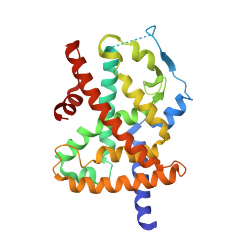
    2P4Y

  • PubMed Abstract: 

    Despite their proven antidiabetic efficacy, widespread use of peroxisome proliferator-activated receptor (PPAR)gamma agonists has been limited by adverse cardiovascular effects. To overcome this shortcoming, selective PPARgamma modulators (SPPARgammaMs) have been identified that have antidiabetic efficacy comparable with full agonists with improved tolerability in preclinical species. The results of structural studies support the proposition that SPPARgammaMs interact with PPARgamma differently from full agonists, thereby providing a physical basis for their novel activities. Herein, we describe a novel PPARgamma ligand, SPPARgammaM2. This compound was a partial agonist in a cell-based transcriptional activity assay, with diminished adipogenic activity and an attenuated gene signature in cultured human adipocytes. X-ray cocrystallography studies demonstrated that, unlike rosiglitazone, SPPARgammaM2 did not interact with the Tyr473 residue located within helix 12 of the ligand binding domain (LBD). Instead, SPPARgammaM2 was found to bind to and activate human PPARgamma in which the Tyr473 residue had been mutated to alanine (hPPARgammaY473A), with potencies similar to those observed with the wild-type receptor (hPPARgammaWT). In additional studies, we found that the intrinsic binding and functional potencies of structurally distinct SPPARgammaMs were not diminished by the Y473A mutation, whereas those of various thiazolidinedione (TZD) and non-TZD PPARgamma full agonists were reduced in a correlative manner. These results directly demonstrate the important role of Tyr473 in mediating the interaction of full agonists but not SPPARgammaMs with the PPARgamma LBD, thereby providing a precise molecular determinant for their differing pharmacologies.


  • Organizational Affiliation
    • Department of Metabolic Disorders, Merck Research Laboratories, 126 E. Lincoln Ave., Rahway, NJ 07065, USA.

Macromolecule Content 

  • Total Structure Weight: 64.46 kDa 
  • Atom Count: 3,973 
  • Modeled Residue Count: 471 
  • Deposited Residue Count: 554 
  • Unique protein chains: 1

Macromolecules

Find similar proteins by:|  3D Structure
Entity ID: 1
MoleculeChains  Sequence LengthOrganismDetailsImage
Peroxisome proliferator-activated receptor gamma
A, B
277Homo sapiensMutation(s): 0 
Gene Names: PPARGNR1C3
UniProt & NIH Common Fund Data Resources
Find proteins for P37231 (Homo sapiens)
Explore P37231 
Go to UniProtKB:  P37231
PHAROS:  P37231
GTEx:  ENSG00000132170 
Entity Groups
Sequence Clusters30% Identity50% Identity70% Identity90% Identity95% Identity100% Identity
UniProt GroupP37231
Sequence Annotations
Expand
Reference Sequence

Small Molecules

Ligands 2 Unique
IDChains Name / Formula / InChI Key2D Diagram3D Interactions
C03

Query on C03


Download:Ideal Coordinates CCD File
C [auth A],
E [auth B]
(2R)-2-(4-CHLORO-3-{[3-(6-METHOXY-1,2-BENZISOXAZOL-3-YL)-2-METHYL-6-(TRIFLUOROMETHOXY)-1H-INDOL-1-YL]METHYL}PHENOXY)PROPANOIC ACID
C28 H22 Cl F3 N2 O6
SCDKVHCGNOYKFK-OAHLLOKOSA-N
TRS

Query on TRS


Download:Ideal Coordinates CCD File
D [auth A]2-AMINO-2-HYDROXYMETHYL-PROPANE-1,3-DIOL
C4 H12 N O3
LENZDBCJOHFCAS-UHFFFAOYSA-O

Experimental Data & Validation

Experimental Data

  • Method: X-RAY DIFFRACTION
  • Resolution: 2.25 Å
  • R-Value Free:  0.274 (Depositor), 0.273 (DCC) 
  • R-Value Work:  0.232 (Depositor), 0.232 (DCC) 
  • R-Value Observed: 0.232 (Depositor) 
Space Group: C 1 2 1
Unit Cell:
Length ( Å )Angle ( ˚ )
a = 92.148α = 90
b = 59.859β = 103.76
c = 118.287γ = 90
Software Package:
Software NamePurpose
CNXrefinement
ADSCdata collection
HKL-2000data reduction
SCALEPACKdata scaling
CNXphasing

Structure Validation

View Full Validation Report


Ligand Structure Quality Assessment 


Entry History 

Deposition Data

Revision History  (Full details and data files)

  • Version 1.0: 2008-01-08
    Type: Initial release
  • Version 1.1: 2011-07-13
    Changes: Version format compliance
  • Version 1.2: 2024-02-21
    Changes: Data collection, Database references, Derived calculations
  • Version 1.3: 2024-04-03
    Changes: Refinement description