2P49

Complex of a camelid single-domain vhh antibody fragment with RNASE A at 1.4A resolution: native mono_1 crystal form


Experimental Data Snapshot

  • Method: X-RAY DIFFRACTION
  • Resolution: 1.38 Å
  • R-Value Free: 0.185 
  • R-Value Work: 0.156 
  • R-Value Observed: 0.157 

Starting Model: experimental
View more details

wwPDB Validation   3D Report Full Report


This is version 1.3 of the entry. See complete history


Literature

Exploring the capacity of minimalist protein interfaces: interface energetics and affinity maturation to picomolar KD of a single-domain antibody with a flat paratope.

Koide, A.Tereshko, V.Uysal, S.Margalef, K.Kossiakoff, A.A.Koide, S.

(2007) J Mol Biol 373: 941-953

  • DOI: https://doi.org/10.1016/j.jmb.2007.08.027
  • Primary Citation of Related Structures:  
    2P49, 2P4A

  • PubMed Abstract: 

    A major architectural class in engineered binding proteins ("antibody mimics") involves the presentation of recognition loops off a single-domain scaffold. This class of binding proteins, both natural and synthetic, has a strong tendency to bind a preformed cleft using a convex binding interface (paratope). To explore their capacity to produce high-affinity interfaces with diverse shape and topography, we examined the interface energetics and explored the affinity limit achievable with a flat paratope. We chose a minimalist paratope limited to two loops found in a natural camelid heavy-chain antibody (VHH) that binds to ribonuclease A. Ala scanning of the VHH revealed only three "hot spot" side chains and additional four residues important for supporting backbone-mediated interactions. The small number of critical residues suggested that this is not an optimized paratope. Using selection from synthetic combinatorial libraries, we enhanced its affinity by >100-fold, resulting in variants with Kd as low as 180 pM with no detectable loss of binding specificity. High-resolution crystal structures revealed that the mutations induced only subtle structural changes but extended the network of interactions. This resulted in an expanded hot spot region including four additional residues located at the periphery of the paratope with a concomitant loss of the so-called "O-ring" arrangement of energetically inert residues. These results suggest that this class of simple, single-domain scaffolds is capable of generating high-performance binding interfaces with diverse shape. More generally, they suggest that highly functional interfaces can be designed without closely mimicking natural interfaces.


  • Organizational Affiliation

    Department of Biochemistry and Molecular Biology, The University of Chicago, Chicago, IL 60637, USA.


Macromolecules
Find similar proteins by:  (by identity cutoff)  |  3D Structure
Entity ID: 1
MoleculeChains Sequence LengthOrganismDetailsImage
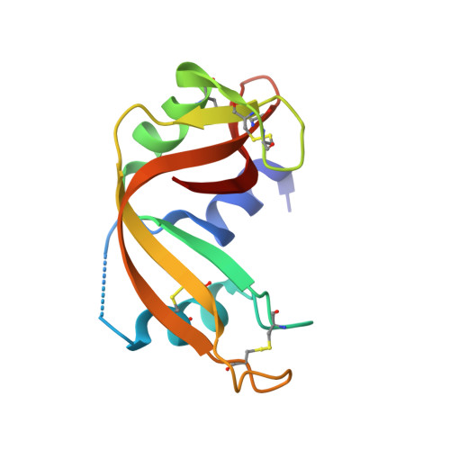
Ribonuclease pancreatic124Bos taurusMutation(s): 0 
EC: 3.1.27.5 (PDB Primary Data), 4.6.1.18 (UniProt)
UniProt
Find proteins for P61823 (Bos taurus)
Explore P61823 
Go to UniProtKB:  P61823
Entity Groups  
Sequence Clusters30% Identity50% Identity70% Identity90% Identity95% Identity100% Identity
UniProt GroupP61823
Sequence Annotations
Expand
  • Reference Sequence
Find similar proteins by:  (by identity cutoff)  |  3D Structure
Entity ID: 2
MoleculeChains Sequence LengthOrganismDetailsImage
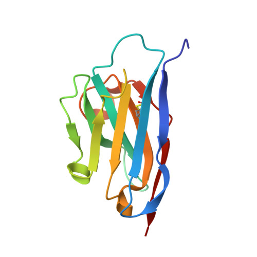
ANTIBODY CAB-RN05123Camelus dromedariusMutation(s): 0 
UniProt
Find proteins for A2KD57 (Lama glama)
Explore A2KD57 
Go to UniProtKB:  A2KD57
Entity Groups  
Sequence Clusters30% Identity50% Identity70% Identity90% Identity95% Identity100% Identity
UniProt GroupA2KD57
Sequence Annotations
Expand
  • Reference Sequence
Small Molecules
Ligands 1 Unique
IDChains Name / Formula / InChI Key2D Diagram3D Interactions
PO4
Query on PO4

Download Ideal Coordinates CCD File 
C [auth A]PHOSPHATE ION
O4 P
NBIIXXVUZAFLBC-UHFFFAOYSA-K
Experimental Data & Validation

Experimental Data

  • Method: X-RAY DIFFRACTION
  • Resolution: 1.38 Å
  • R-Value Free: 0.185 
  • R-Value Work: 0.156 
  • R-Value Observed: 0.157 
  • Space Group: P 1 21 1
Unit Cell:
Length ( Å )Angle ( ˚ )
a = 40.13α = 90
b = 54.394β = 108.49
c = 48.044γ = 90
Software Package:
Software NamePurpose
REFMACrefinement
HKL-3000data collection
HKL-3000data reduction
HKL-3000data scaling
MOLREPphasing

Structure Validation

View Full Validation Report


Entry History 

Deposition Data

Revision History  (Full details and data files)

  • Version 1.0: 2007-08-28
    Type: Initial release
  • Version 1.1: 2011-07-13
    Changes: Advisory, Version format compliance
  • Version 1.2: 2023-08-30
    Changes: Data collection, Database references, Derived calculations, Refinement description
  • Version 1.3: 2024-11-13
    Changes: Structure summary