2P3O | pdb_00002p3o

Crystal Structure of Dengue Methyltransferase in Complex with 7MeGpppA and S-Adenosyl-L-homocysteine


Experimental Data Snapshot

  • Method: X-RAY DIFFRACTION
  • Resolution: 2.76 Å
  • R-Value Free: 
    0.244 (Depositor), 0.231 (DCC) 
  • R-Value Work: 
    0.195 (Depositor) 
  • R-Value Observed: 
    0.198 (Depositor) 

Starting Model: experimental
View more details

wwPDB Validation 3D Report Full Report

Validation slider image for 2P3O

Ligand Structure Quality Assessment 


This is version 1.2 of the entry. See complete history

Literature

Structural and functional analysis of methylation and 5'-RNA sequence requirements of short capped RNAs by the methyltransferase domain of dengue virus NS5

Egloff, M.P.Decroly, E.Malet, H.Selisko, B.Benarroch, D.Ferron, F.Canard, B.

(2007) J Mol Biology 372: 723-736

  • DOI: https://doi.org/10.1016/j.jmb.2007.07.005
  • Primary Citation Related Structures: 
    2P3L, 2P3O, 2P3Q, 2P40, 2P41

  • PubMed Abstract: 

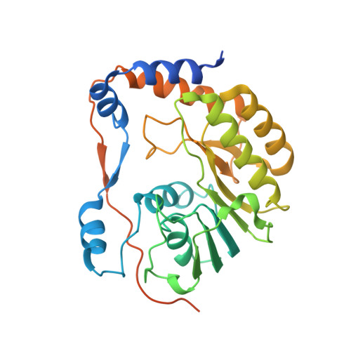
    The N-terminal 33 kDa domain of non-structural protein 5 (NS5) of dengue virus (DV), named NS5MTase(DV), is involved in two of four steps required for the formation of the viral mRNA cap (7Me)GpppA(2'OMe), the guanine-N7 and the adenosine-2'O methylation. Its S-adenosyl-l-methionine (AdoMet) dependent 2'O-methyltransferase (MTase) activity has been shown on capped (7Me+/-)GpppAC(n) RNAs. Here we report structural and binding studies using cap analogues and capped RNAs. We have solved five crystal structures at 1.8 A to 2.8 A resolution of NS5MTase(DV) in complex with cap analogues and the co-product of methylation S-adenosyl-l-homocysteine (AdoHcy). The cap analogues can adopt several conformations. The guanosine moiety of all cap analogues occupies a GTP-binding site identified earlier, indicating that GTP and cap share the same binding site. Accordingly, we show that binding of (7Me)GpppAC(4) and (7Me)GpppAC(5) RNAs is inhibited in the presence of GTP, (7Me)GTP and (7Me)GpppA but not by ATP. This particular position of the cap is in accordance with the 2'O-methylation step. A model was generated of a ternary 2'O-methylation complex of NS5MTase(DV), (7Me)GpppA and AdoMet. RNA-binding increased when (7Me+/-)GpppAGC(n-1) starting with the consensus sequence GpppAG, was used instead of (7Me+/-)GpppAC(n). In the NS5MTase(DV)-GpppA complex the cap analogue adopts a folded, stacked conformation uniquely possible when adenine is the first transcribed nucleotide at the 5' end of nascent RNA, as it is the case in all flaviviruses. This conformation cannot be a functional intermediate of methylation, since both the guanine-N7 and adenosine-2'O positions are too far away from AdoMet. We hypothesize that this conformation mimics the reaction product of a yet-to-be-demonstrated guanylyltransferase activity. A putative Flavivirus RNA capping pathway is proposed combining the different steps where the NS5MTase domain is involved.


  • Organizational Affiliation
    • Architecture et Fonction des Macromolécules Biologiques, CNRS and Universités d'Aix-Marseille I et II, UMR 6098, ESIL Case 925, 13288 Marseille, France.

Macromolecule Content 

  • Total Structure Weight: 36.63 kDa 
  • Atom Count: 2,184 
  • Modeled Residue Count: 258 
  • Deposited Residue Count: 305 
  • Unique protein chains: 1

Macromolecules

Find similar proteins by:|  3D Structure
Entity ID: 1
MoleculeChains  Sequence LengthOrganismDetailsImage
type II methyltransferase305dengue virus type 2Mutation(s): 0 
Gene Names: NSP5
EC: 2.7.7.48
UniProt
Find proteins for P12823 (Dengue virus type 2 (strain Puerto Rico/PR159-S1/1969))
Explore P12823 
Go to UniProtKB:  P12823
Entity Groups
Sequence Clusters30% Identity50% Identity70% Identity90% Identity95% Identity100% Identity
UniProt GroupP12823
Sequence Annotations
Expand
Reference Sequence

Small Molecules

Ligands 4 Unique
IDChains Name / Formula / InChI Key2D Diagram3D Interactions
GTA

Query on GTA


Download:Ideal Coordinates CCD File
M [auth A]P1-7-METHYLGUANOSINE-P3-ADENOSINE-5',5'-TRIPHOSPHATE
C21 H30 N10 O17 P3
QQOHNVHGNZYSBP-XPWFQUROSA-O
SAH

Query on SAH


Download:Ideal Coordinates CCD File
K [auth A]S-ADENOSYL-L-HOMOCYSTEINE
C14 H20 N6 O5 S
ZJUKTBDSGOFHSH-WFMPWKQPSA-N
CIT

Query on CIT


Download:Ideal Coordinates CCD File
L [auth A]CITRIC ACID
C6 H8 O7
KRKNYBCHXYNGOX-UHFFFAOYSA-N
SO4

Query on SO4


Download:Ideal Coordinates CCD File
B [auth A]
C [auth A]
D [auth A]
E [auth A]
F [auth A]
B [auth A],
C [auth A],
D [auth A],
E [auth A],
F [auth A],
G [auth A],
H [auth A],
I [auth A],
J [auth A]
SULFATE ION
O4 S
QAOWNCQODCNURD-UHFFFAOYSA-L

Experimental Data & Validation

Experimental Data

  • Method: X-RAY DIFFRACTION
  • Resolution: 2.76 Å
  • R-Value Free:  0.244 (Depositor), 0.231 (DCC) 
  • R-Value Work:  0.195 (Depositor) 
  • R-Value Observed: 0.198 (Depositor) 
Space Group: P 31 2 1
Unit Cell:
Length ( Å )Angle ( ˚ )
a = 111.56α = 90
b = 111.56β = 90
c = 56.33γ = 120
Software Package:
Software NamePurpose
ADSCdata collection
AMoREphasing
REFMACrefinement
MOSFLMdata reduction
SCALAdata scaling

Structure Validation

View Full Validation Report


Ligand Structure Quality Assessment 


Entry History 

Deposition Data

Revision History  (Full details and data files)

  • Version 1.0: 2007-08-28
    Type: Initial release
  • Version 1.1: 2011-07-13
    Changes: Derived calculations, Version format compliance
  • Version 1.2: 2023-08-30
    Changes: Data collection, Database references, Derived calculations, Refinement description