2P3G | pdb_00002p3g

Crystal structure of a pyrrolopyridine inhibitor bound to MAPKAP Kinase-2


Experimental Data Snapshot

  • Method: X-RAY DIFFRACTION
  • Resolution: 3.80 Å
  • R-Value Free: 
    0.374 (Depositor), 0.348 (DCC) 
  • R-Value Work: 
    0.300 (Depositor), 0.279 (DCC) 
  • R-Value Observed: 
    0.303 (Depositor) 

Starting Model: experimental
View more details

wwPDB Validation 3D Report Full Report

Validation slider image for 2P3G

Ligand Structure Quality Assessment 


This is version 1.5 of the entry. See complete history

Literature

Pyrrolopyridine Inhibitors of Mitogen-Activated Protein Kinase-Activated Protein Kinase 2 (MK-2).

Anderson, D.R.Meyers, M.J.Vernier, W.F.Mahoney, M.W.Kurumbail, R.G.Caspers, N.Poda, G.I.Schindler, J.F.Reitz, D.B.Mourey, R.J.

(2007) J Med Chem 50: 2647-2654

  • DOI: https://doi.org/10.1021/jm0611004
  • Primary Citation Related Structures: 
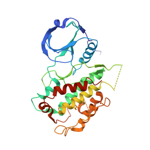
    2P3G

  • PubMed Abstract: 

    A new class of potent kinase inhibitors selective for mitogen-activated protein kinase-activated protein kinase 2 (MAPKAP-K2 or MK-2) for the treatment of rheumatoid arthritis has been prepared and evaluated. These inhibitors have IC50 values as low as 10 nM against the target and have good selectivity profiles against a number of kinases including CDK2, ERK, JNK, and p38. These MK-2 inhibitors have been shown to suppress TNFalpha production in U397 cells and to be efficacious in an acute inflammation model. The structure-activity relationships of this series, the selectivity for MK-2 and their activity in both in vitro and in vivo models are discussed. The observed selectivity is discussed with the aid of an MK-2/inhibitor crystal structure.


  • Organizational Affiliation
    • Pfizer Global Research and Development, St. Louis Laboratories, 700 Chesterfield Parkway W, Chesterfield, Missouri 63017, USA. david.r.anderson@pfizer.com

Macromolecule Content 

  • Total Structure Weight: 38.21 kDa 
  • Atom Count: 2,321 
  • Modeled Residue Count: 282 
  • Deposited Residue Count: 327 
  • Unique protein chains: 1

Macromolecules

Find similar proteins by:|  3D Structure
Entity ID: 1
MoleculeChains  Sequence LengthOrganismDetailsImage
MAP kinase-activated protein kinase 2A [auth X]327Homo sapiensMutation(s): 0 
Gene Names: MAPKAPK2
EC: 2.7.11.1
UniProt & NIH Common Fund Data Resources
Find proteins for P49137 (Homo sapiens)
Explore P49137 
Go to UniProtKB:  P49137
PHAROS:  P49137
GTEx:  ENSG00000162889 
Entity Groups
Sequence Clusters30% Identity50% Identity70% Identity90% Identity95% Identity100% Identity
UniProt GroupP49137
Sequence Annotations
Expand
Reference Sequence

Small Molecules

Ligands 1 Unique
IDChains Name / Formula / InChI Key2D Diagram3D Interactions
F10

Query on F10


Download:Ideal Coordinates CCD File
B [auth X]2-[2-(2-FLUOROPHENYL)PYRIDIN-4-YL]-1,5,6,7-TETRAHYDRO-4H-PYRROLO[3,2-C]PYRIDIN-4-ONE
C18 H14 F N3 O
XJJYJNMNYDNXNO-UHFFFAOYSA-N

Experimental Data & Validation

Experimental Data

  • Method: X-RAY DIFFRACTION
  • Resolution: 3.80 Å
  • R-Value Free:  0.374 (Depositor), 0.348 (DCC) 
  • R-Value Work:  0.300 (Depositor), 0.279 (DCC) 
  • R-Value Observed: 0.303 (Depositor) 
Space Group: F 41 3 2
Unit Cell:
Length ( Å )Angle ( ˚ )
a = 253.941α = 90
b = 253.941β = 90
c = 253.941γ = 90
Software Package:
Software NamePurpose
DENZOdata reduction
SCALEPACKdata scaling
REFMACrefinement
PDB_EXTRACTdata extraction
ADSCdata collection
HKL-2000data reduction
X-PLORphasing

Structure Validation

View Full Validation Report


Ligand Structure Quality Assessment 


Entry History 

Deposition Data

Revision History  (Full details and data files)

  • Version 1.0: 2007-06-12
    Type: Initial release
  • Version 1.1: 2008-03-05
    Changes: Version format compliance
  • Version 1.2: 2011-07-13
    Changes: Version format compliance
  • Version 1.3: 2017-10-18
    Changes: Refinement description
  • Version 1.4: 2024-02-21
    Changes: Data collection, Database references, Derived calculations
  • Version 1.5: 2024-04-03
    Changes: Refinement description