2P1Y | pdb_00002p1y

1.B2.D9, a bispecific alpha/beta TCR


Experimental Data Snapshot

  • Method: X-RAY DIFFRACTION
  • Resolution: 2.42 Å
  • R-Value Free: 
    0.254 (Depositor), 0.218 (DCC) 
  • R-Value Work: 
    0.181 (Depositor), 0.188 (DCC) 
  • R-Value Observed: 
    0.185 (Depositor) 

Starting Model: experimental
View more details

wwPDB Validation 3D Report Full Report

Validation slider image for 2P1Y

This is version 1.3 of the entry. See complete history

Literature

A new twist in TCR diversity revealed by a forbidden alphabeta TCR.

McBeth, C.Seamons, A.Pizarro, J.C.Fleishman, S.J.Baker, D.Kortemme, T.Goverman, J.M.Strong, R.K.

(2008) J Mol Biology 375: 1306-1319

  • DOI: https://doi.org/10.1016/j.jmb.2007.11.020
  • Primary Citation Related Structures: 
    2P1Y, 2P24

  • PubMed Abstract: 

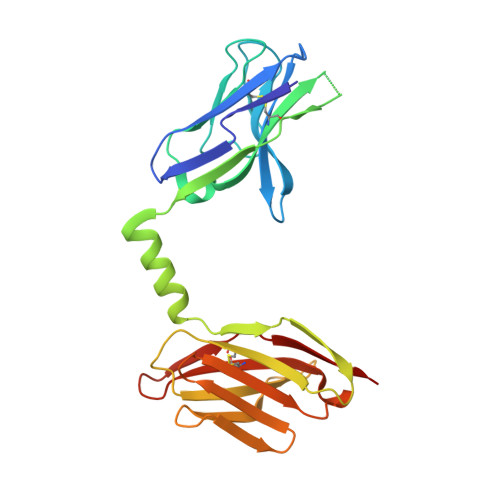
    We report crystal structures of a negatively selected T cell receptor (TCR) that recognizes two I-A(u)-restricted myelin basic protein peptides and one of its peptide/major histocompatibility complex (pMHC) ligands. Unusual complementarity-determining region (CDR) structural features revealed by our analyses identify a previously unrecognized mechanism by which the highly variable CDR3 regions define ligand specificity. In addition to the pMHC contact residues contributed by CDR3, the CDR3 residues buried deep within the V alpha/V beta interface exert indirect effects on recognition by influencing the V alpha/V beta interdomain angle. This phenomenon represents an additional mechanism for increasing the potential diversity of the TCR repertoire. Both the direct and indirect effects exerted by CDR residues can impact global TCR/MHC docking. Analysis of the available TCR structures in light of these results highlights the significance of the V alpha/V beta interdomain angle in determining specificity and indicates that TCR/pMHC interface features do not distinguish autoimmune from non-autoimmune class II-restricted TCRs.


  • Organizational Affiliation
    • Division of Basic Sciences, Fred Hutchinson Cancer Research Center, Seattle, WA 98109, USA.

Macromolecule Content 

  • Total Structure Weight: 105.07 kDa 
  • Atom Count: 7,017 
  • Modeled Residue Count: 896 
  • Deposited Residue Count: 952 
  • Unique protein chains: 1

Macromolecules

Find similar proteins by:|  3D Structure
Entity ID: 1
MoleculeChains  Sequence LengthOrganismDetailsImage
bispecific alpha/beta TCRA,
B [auth C],
C [auth E],
D [auth G]
238Mus musculusMutation(s): 0 
Gene Names: Valpha2.3/Vbeta8.2
UniProt
Find proteins for Q5R1B3 (Mus musculus)
Explore Q5R1B3 
Go to UniProtKB:  Q5R1B3
Entity Groups
Sequence Clusters30% Identity50% Identity70% Identity90% Identity95% Identity100% Identity
UniProt GroupQ5R1B3
Sequence Annotations
Expand
Reference Sequence

Experimental Data & Validation

Experimental Data

  • Method: X-RAY DIFFRACTION
  • Resolution: 2.42 Å
  • R-Value Free:  0.254 (Depositor), 0.218 (DCC) 
  • R-Value Work:  0.181 (Depositor), 0.188 (DCC) 
  • R-Value Observed: 0.185 (Depositor) 
Space Group: P 1 21 1
Unit Cell:
Length ( Å )Angle ( ˚ )
a = 71.681α = 90
b = 105.477β = 116.04
c = 81.531γ = 90
Software Package:
Software NamePurpose
REFMACrefinement
HKL-2000data collection
DENZOdata reduction
SCALEPACKdata scaling
AMoREphasing

Structure Validation

View Full Validation Report


Entry History 

Deposition Data

Revision History  (Full details and data files)

  • Version 1.0: 2008-02-05
    Type: Initial release
  • Version 1.1: 2011-07-13
    Changes: Version format compliance
  • Version 1.2: 2023-08-30
    Changes: Data collection, Database references, Refinement description
  • Version 1.3: 2024-11-20
    Changes: Structure summary