2OXP | pdb_00002oxp

Crystal Structure of Staphylococcal Nuclease mutant V66D/P117G/H124L/S128A


Experimental Data Snapshot

  • Method: X-RAY DIFFRACTION
  • Resolution: 2.00 Å
  • R-Value Free: 
    0.234 (Depositor) 
  • R-Value Work: 
    0.207 (Depositor) 

wwPDB Validation 3D Report Full Report

Validation slider image for 2OXP

This is version 1.4 of the entry. See complete history

Literature

High apparent dielectric constant inside a protein reflects structural reorganization coupled to the ionization of an internal asp

Karp, D.A.Gittis, A.G.Stahley, M.R.Fitch, C.A.Stites, W.E.Garcia-Moreno, E.B.

(2007) Biophys J 92: 2041-2053

  • DOI: https://doi.org/10.1529/biophysj.106.090266
  • Primary Citation Related Structures: 
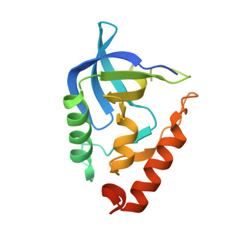
    2OXP

  • PubMed Abstract: 

    The dielectric properties of proteins are poorly understood and difficult to describe quantitatively. This limits the accuracy of methods for structure-based calculation of electrostatic energies and pK(a) values. The pK(a) values of many internal groups report apparent protein dielectric constants of 10 or higher. These values are substantially higher than the dielectric constants of 2-4 measured experimentally with dry proteins. The structural origins of these high apparent dielectric constants are not well understood. Here we report on structural and equilibrium thermodynamic studies of the effects of pH on the V66D variant of staphylococcal nuclease. In a crystal structure of this protein the neutral side chain of Asp-66 is buried in the hydrophobic core of the protein and hydrated by internal water molecules. Asp-66 titrates with a pK(a) value near 9. A decrease in the far UV-CD signal was observed, concomitant with ionization of this aspartic acid, and consistent with the loss of 1.5 turns of alpha-helix. These data suggest that the protein dielectric constant needed to reproduce the pK(a) value of Asp-66 with continuum electrostatics calculations is high because the dielectric constant has to capture, implicitly, the energetic consequences of the structural reorganization that are not treated explicitly in continuum calculations with static structures.


  • Organizational Affiliation
    • Department of Biophysics, Johns Hopkins University, Baltimore, Maryland, USA.

Macromolecule Content 

  • Total Structure Weight: 17.18 kDa 
  • Atom Count: 1,131 
  • Modeled Residue Count: 129 
  • Deposited Residue Count: 149 
  • Unique protein chains: 1

Macromolecules

Find similar proteins by:|  3D Structure
Entity ID: 1
MoleculeChains  Sequence LengthOrganismDetailsImage
Thermonuclease149Staphylococcus aureusMutation(s): 3 
EC: 3.1.31.1
UniProt
Find proteins for Q8NXI6 (Staphylococcus aureus (strain MW2))
Explore Q8NXI6 
Go to UniProtKB:  Q8NXI6
Entity Groups
Sequence Clusters30% Identity50% Identity70% Identity90% Identity95% Identity100% Identity
UniProt GroupQ8NXI6
Sequence Annotations
Expand
Reference Sequence

Small Molecules

Ligands 1 Unique
IDChains Name / Formula / InChI Key2D Diagram3D Interactions
THP

Query on THP


Download:Ideal Coordinates CCD File
B [auth A]THYMIDINE-3',5'-DIPHOSPHATE
C10 H16 N2 O11 P2
CSNCBOPUCJOHLS-XLPZGREQSA-N

Experimental Data & Validation

Experimental Data

  • Method: X-RAY DIFFRACTION
  • Resolution: 2.00 Å
  • R-Value Free:  0.234 (Depositor) 
  • R-Value Work:  0.207 (Depositor) 
Space Group: P 41
Unit Cell:
Length ( Å )Angle ( ˚ )
a = 47.579α = 90
b = 47.579β = 90
c = 62.791γ = 90
Software Package:
Software NamePurpose
CNSrefinement
PDB_EXTRACTdata extraction
HKL-2000data collection
DENZOdata reduction
SCALEPACKdata scaling
CNSphasing

Structure Validation

View Full Validation Report


Entry History 

Deposition Data

Revision History  (Full details and data files)

  • Version 1.0: 2007-03-20
    Type: Initial release
  • Version 1.1: 2008-05-01
    Changes: Version format compliance
  • Version 1.2: 2011-07-13
    Changes: Version format compliance
  • Version 1.3: 2021-10-20
    Changes: Database references, Derived calculations
  • Version 1.4: 2024-02-21
    Changes: Data collection