2OWO | pdb_00002owo

Last Stop on the Road to Repair: Structure of E.coli DNA Ligase Bound to Nicked DNA-Adenylate


Experimental Data Snapshot

  • Method: X-RAY DIFFRACTION
  • Resolution: 2.30 Å
  • R-Value Free: 
    0.254 (Depositor), 0.220 (DCC) 
  • R-Value Work: 
    0.217 (Depositor), 0.222 (DCC) 

Starting Models: experimental
View more details

wwPDB Validation 3D Report Full Report

Validation slider image for 2OWO

Ligand Structure Quality Assessment 


This is version 1.4 of the entry. See complete history

Literature

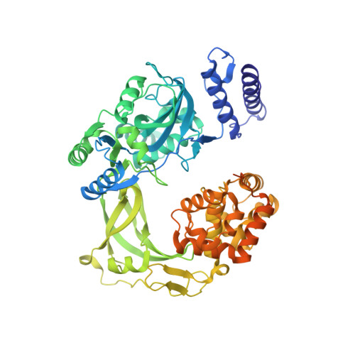
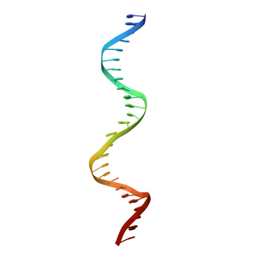
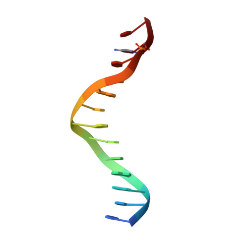
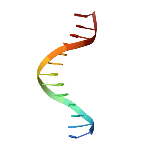
Last Stop on the Road to Repair: Structure of E. coli DNA Ligase Bound to Nicked DNA-Adenylate.

Nandakumar, J.Nair, P.A.Shuman, S.

(2007) Mol Cell 26: 257-271

  • DOI: https://doi.org/10.1016/j.molcel.2007.02.026
  • Primary Citation Related Structures: 
    2OWO

  • PubMed Abstract: 

    NAD(+)-dependent DNA ligases (LigA) are ubiquitous in bacteria and essential for growth. Their distinctive substrate specificity and domain organization vis-a-vis human ATP-dependent ligases make them outstanding targets for anti-infective drug discovery. We report here the 2.3 A crystal structure of Escherichia coli LigA bound to an adenylylated nick, which captures LigA in a state poised for strand closure and reveals the basis for nick recognition. LigA envelopes the DNA within a protein clamp. Large protein domain movements and remodeling of the active site orchestrate progression through the three chemical steps of the ligation reaction. The structure inspires a strategy for inhibitor design.


  • Organizational Affiliation
    • Molecular Biology Program, Sloan-Kettering Institute, New York, NY 10021, USA.

Macromolecule Content 

  • Total Structure Weight: 90.46 kDa 
  • Atom Count: 5,991 
  • Modeled Residue Count: 638 
  • Deposited Residue Count: 723 
  • Unique protein chains: 1
  • Unique nucleic acid chains: 3

Macromolecules


Find similar proteins by:|  3D Structure
Entity ID: 4
MoleculeChains  Sequence LengthOrganismDetailsImage
DNA ligaseD [auth A]671Escherichia coli K-12Mutation(s): 0 
Gene Names: ligA
EC: 6.5.1.2
UniProt
Find proteins for P15042 (Escherichia coli (strain K12))
Explore P15042 
Go to UniProtKB:  P15042
Entity Groups
Sequence Clusters30% Identity50% Identity70% Identity90% Identity95% Identity100% Identity
UniProt GroupP15042
Sequence Annotations
Expand
Reference Sequence
Find similar nucleic acids by:  (by identity cutoff) 
Entity ID: 1
MoleculeChains LengthOrganismImage
26-MERA [auth B]26N/A
Sequence Annotations
Expand
Reference Sequence
Find similar nucleic acids by:  Sequence
Entity ID: 2
MoleculeChains LengthOrganismImage
5'-D(*AP*CP*AP*AP*TP*TP*GP*CP*GP*AP*CP*(OMC)P*C)-3'B [auth C]13N/A
Sequence Annotations
Expand
Reference Sequence
Find similar nucleic acids by:  Sequence
Entity ID: 3
MoleculeChains LengthOrganismImage
5'-D(*CP*AP*CP*TP*AP*TP*CP*GP*GP*AP*AP*TP*G)-3'C [auth D]13N/A
Sequence Annotations
Expand
Reference Sequence

Experimental Data & Validation

Experimental Data

  • Method: X-RAY DIFFRACTION
  • Resolution: 2.30 Å
  • R-Value Free:  0.254 (Depositor), 0.220 (DCC) 
  • R-Value Work:  0.217 (Depositor), 0.222 (DCC) 
Space Group: C 1 2 1
Unit Cell:
Length ( Å )Angle ( ˚ )
a = 110.441α = 90
b = 99.273β = 105.33
c = 86.245γ = 90
Software Package:
Software NamePurpose
DENZOdata reduction
SCALEPACKdata scaling
CNSrefinement
PDB_EXTRACTdata extraction
ADSCdata collection
MOLREPphasing

Structure Validation

View Full Validation Report


Ligand Structure Quality Assessment 


Entry History 

Deposition Data

Revision History  (Full details and data files)

  • Version 1.0: 2007-05-15
    Type: Initial release
  • Version 1.1: 2008-05-01
    Changes: Version format compliance
  • Version 1.2: 2011-07-13
    Changes: Source and taxonomy, Version format compliance
  • Version 1.3: 2017-10-18
    Changes: Refinement description
  • Version 1.4: 2023-08-30
    Changes: Data collection, Database references, Derived calculations, Refinement description