2OR8 | pdb_00002or8

Tim-1


Experimental Data Snapshot

  • Method: X-RAY DIFFRACTION
  • Resolution: 2.50 Å
  • R-Value Free: 
    0.278 (Depositor) 
  • R-Value Work: 
    0.232 (Depositor), 0.240 (DCC) 
  • R-Value Observed: 
    0.241 (Depositor) 

Starting Model: experimental
View more details

wwPDB Validation 3D Report Full Report

Validation slider image for 2OR8

This is version 1.4 of the entry. See complete history

Literature

Structures of T Cell Immunoglobulin Mucin Receptors 1 and 2 Reveal Mechanisms for Regulation of Immune Responses by the TIM Receptor Family.

Santiago, C.Ballesteros, A.Tami, C.Martinez-Munoz, L.Kaplan, G.G.Casasnovas, J.M.

(2007) Immunity 26: 299-310

  • DOI: https://doi.org/10.1016/j.immuni.2007.01.014
  • Primary Citation Related Structures: 
    2OR7, 2OR8

  • PubMed Abstract: 

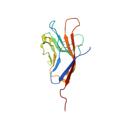
    The T cell immunoglobulin mucin (TIM) receptors are involved in the regulation of immune responses, autoimmunity, and allergy. Structures of the N-terminal ligand binding domain of the murine mTIM-1 and mTIM-2 receptors revealed an immunoglobulin (Ig) fold, with four Cys residues bridging a distinctive CC' loop to the GFC beta-sheet. The structures showed two ligand-recognition modes in the TIM family. The mTIM-1 structure identified a homophilic TIM-TIM adhesion interaction, whereas the mTIM-2 domain formed a dimer that prevented homophilic binding. Biochemical, mutational, and cell adhesion analyses confirmed the divergent ligand-binding modes revealed by the structures. Structural features characteristic of mTIM-1 appear conserved in human TIM-1, which also mediated homophilic interactions. The extracellular mucin domain enhanced binding through the Ig domain, modulating TIM receptor functions. These results explain the divergent immune functions described for the murine receptors and the role of TIM-1 as a cell adhesion receptor in renal regeneration and cancer.


  • Organizational Affiliation
    • Centro Nacional de Biotecnologia, CSIC, Campus Universidad Autónoma, 28049 Madrid, Spain.

Macromolecule Content 

  • Total Structure Weight: 26.02 kDa 
  • Atom Count: 1,926 
  • Modeled Residue Count: 232 
  • Deposited Residue Count: 232 
  • Unique protein chains: 1

Macromolecules

Find similar proteins by:|  3D Structure
Entity ID: 1
MoleculeChains  Sequence LengthOrganismDetailsImage
Hepatitis A virus cellular receptor 1 homolog
A, B
116Mus musculusMutation(s): 0 
Gene Names: Havcr1Tim1Timd1
Membrane Entity: Yes 
UniProt
Find proteins for Q5QNS5 (Mus musculus)
Explore Q5QNS5 
Go to UniProtKB:  Q5QNS5
Entity Groups
Sequence Clusters30% Identity50% Identity70% Identity90% Identity95% Identity100% Identity
UniProt GroupQ5QNS5
Sequence Annotations
Expand
Reference Sequence

Small Molecules

Ligands 1 Unique
IDChains Name / Formula / InChI Key2D Diagram3D Interactions
ACT

Query on ACT


Download:Ideal Coordinates CCD File
C [auth A]ACETATE ION
C2 H3 O2
QTBSBXVTEAMEQO-UHFFFAOYSA-M

Experimental Data & Validation

Experimental Data

  • Method: X-RAY DIFFRACTION
  • Resolution: 2.50 Å
  • R-Value Free:  0.278 (Depositor) 
  • R-Value Work:  0.232 (Depositor), 0.240 (DCC) 
  • R-Value Observed: 0.241 (Depositor) 
Space Group: P 21 21 21
Unit Cell:
Length ( Å )Angle ( ˚ )
a = 44.49α = 90
b = 55.74β = 90
c = 75.53γ = 90
Software Package:
Software NamePurpose
PDCdata collection
PHASERphasing
CNSrefinement
XDSdata reduction
CCP4data scaling

Structure Validation

View Full Validation Report


Entry History 

Deposition Data

Revision History  (Full details and data files)

  • Version 1.0: 2007-04-03
    Type: Initial release
  • Version 1.1: 2008-05-01
    Changes: Version format compliance
  • Version 1.2: 2011-07-13
    Changes: Version format compliance
  • Version 1.3: 2024-04-03
    Changes: Data collection, Database references, Derived calculations, Refinement description
  • Version 1.4: 2024-10-16
    Changes: Structure summary