2OOV | pdb_00002oov

Crystal Structure of Hansenula polymorpha amine oxidase to 1.7 Angstroms


Experimental Data Snapshot

  • Method: X-RAY DIFFRACTION
  • Resolution: 1.70 Å
  • R-Value Free: 
    0.178 (Depositor), 0.180 (DCC) 
  • R-Value Work: 
    0.147 (Depositor), 0.150 (DCC) 
  • R-Value Observed: 
    0.149 (Depositor) 

Starting Model: experimental
View more details

wwPDB Validation 3D Report Full Report

Validation slider image for 2OOV

This is version 1.5 of the entry. See complete history

Literature

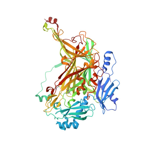
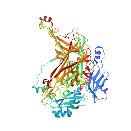
Exploring molecular oxygen pathways in Hansenula polymorpha copper-containing amine oxidase

Johnson, B.J.Cohen, J.Welford, R.W.Pearson, A.R.Schulten, K.Klinman, J.P.Wilmot, C.M.

(2007) J Biological Chem 282: 17767-17776

  • DOI: https://doi.org/10.1074/jbc.M701308200
  • Primary Citation Related Structures: 
    2OOV, 2OQE

  • PubMed Abstract: 

    The accessibility of large substrates to buried enzymatic active sites is dependent upon the utilization of proteinaceous channels. The necessity of these channels in the case of small substrates is questionable because diffusion through the protein matrix is often assumed. Copper amine oxidases contain a buried protein-derived quinone cofactor and a mononuclear copper center that catalyze the conversion of two substrates, primary amines and molecular oxygen, to aldehydes and hydrogen peroxide, respectively. The nature of molecular oxygen migration to the active site in the enzyme from Hansenula polymorpha is explored using a combination of kinetic, x-ray crystallographic, and computational approaches. A crystal structure of H. polymorpha amine oxidase in complex with xenon gas, which serves as an experimental probe for molecular oxygen binding sites, reveals buried regions of the enzyme suitable for transient molecular oxygen occupation. Calculated O(2) free energy maps using copper amine oxidase crystal structures in the absence of xenon correspond well with later experimentally observed xenon sites in these systems, and allow the visualization of O(2) migration routes of differing probabilities within the protein matrix. Site-directed mutagenesis designed to block individual routes has little effect on overall k(cat)/K(m) (O(2)), supporting multiple dynamic pathways for molecular oxygen to reach the active site.


  • Organizational Affiliation
    • Department of Biochemistry, Molecular Biology and Biophysics, University of Minnesota, Minneapolis, Minnesota 55455, USA.

Macromolecule Content 

  • Total Structure Weight: 447.55 kDa 
  • Atom Count: 36,770 
  • Modeled Residue Count: 3,930 
  • Deposited Residue Count: 3,960 
  • Unique protein chains: 2

Macromolecules

Find similar proteins by:|  3D Structure
Entity ID: 1
MoleculeChains  Sequence LengthOrganismDetailsImage
Peroxisomal copper amine oxidase
A, B
660Ogataea angustaMutation(s): 0 
Gene Names: AMO
EC: 1.4.3.6 (PDB Primary Data), 1.4.3.21 (UniProt)
UniProt
Find proteins for P12807 (Pichia angusta)
Explore P12807 
Go to UniProtKB:  P12807
Entity Groups
Sequence Clusters30% Identity50% Identity70% Identity90% Identity95% Identity100% Identity
UniProt GroupP12807
Sequence Annotations
Expand
Reference Sequence
Find similar proteins by:|  3D Structure
Entity ID: 2
MoleculeChains  Sequence LengthOrganismDetailsImage
Peroxisomal copper amine oxidase
C, D, E, F
660Ogataea angustaMutation(s): 0 
Gene Names: AMO
EC: 1.4.3.6 (PDB Primary Data), 1.4.3.21 (UniProt)
UniProt
Find proteins for P12807 (Pichia angusta)
Explore P12807 
Go to UniProtKB:  P12807
Entity Groups
Sequence Clusters30% Identity50% Identity70% Identity90% Identity95% Identity100% Identity
UniProt GroupP12807
Sequence Annotations
Expand
Reference Sequence

Small Molecules

Ligands 3 Unique
IDChains Name / Formula / InChI Key2D Diagram3D Interactions
PO4

Query on PO4


Download:Ideal Coordinates CCD File
LA [auth F],
V [auth C]
PHOSPHATE ION
O4 P
NBIIXXVUZAFLBC-UHFFFAOYSA-K
GOL

Query on GOL


Download:Ideal Coordinates CCD File
BA [auth D]
CA [auth D]
EA [auth E]
FA [auth E]
GA [auth E]
BA [auth D],
CA [auth D],
EA [auth E],
FA [auth E],
GA [auth E],
H [auth A],
HA [auth E],
I [auth A],
IA [auth E],
J [auth A],
JA [auth E],
K [auth A],
L [auth A],
M [auth A],
MA [auth F],
NA [auth F],
O [auth B],
OA [auth F],
P [auth B],
PA [auth F],
Q [auth B],
R [auth B],
S [auth B],
T [auth B],
W [auth C],
X [auth C],
Y [auth C],
Z [auth C]
GLYCEROL
C3 H8 O3
PEDCQBHIVMGVHV-UHFFFAOYSA-N
CU

Query on CU


Download:Ideal Coordinates CCD File
AA [auth D]
DA [auth E]
G [auth A]
KA [auth F]
N [auth B]
AA [auth D],
DA [auth E],
G [auth A],
KA [auth F],
N [auth B],
U [auth C]
COPPER (II) ION
Cu
JPVYNHNXODAKFH-UHFFFAOYSA-N
Modified Residues  2 Unique
IDChains TypeFormula2D DiagramParent
SME
Query on SME
A, B
L-PEPTIDE LINKINGC5 H11 N O3 SMET
TPQ
Query on TPQ
A, B
L-PEPTIDE LINKINGC9 H9 N O5TYR

Experimental Data & Validation

Experimental Data

  • Method: X-RAY DIFFRACTION
  • Resolution: 1.70 Å
  • R-Value Free:  0.178 (Depositor), 0.180 (DCC) 
  • R-Value Work:  0.147 (Depositor), 0.150 (DCC) 
  • R-Value Observed: 0.149 (Depositor) 
Space Group: P 1 21 1
Unit Cell:
Length ( Å )Angle ( ˚ )
a = 104.146α = 90
b = 223.082β = 95.77
c = 104.248γ = 90
Software Package:
Software NamePurpose
DENZOdata reduction
SCALEPACKdata scaling
MOLREPphasing
REFMACrefinement
PDB_EXTRACTdata extraction
HKL-3000data collection
HKL-3000data reduction

Structure Validation

View Full Validation Report


Entry History 

Deposition Data

Revision History  (Full details and data files)

  • Version 1.0: 2007-04-10
    Type: Initial release
  • Version 1.1: 2008-05-01
    Changes: Version format compliance
  • Version 1.2: 2011-07-13
    Changes: Derived calculations, Non-polymer description, Version format compliance
  • Version 1.3: 2012-04-18
    Changes: Derived calculations
  • Version 1.4: 2017-10-18
    Changes: Refinement description
  • Version 1.5: 2023-08-30
    Changes: Data collection, Database references, Derived calculations, Refinement description