2OKD | pdb_00002okd

High Resolution Crystal Structures of Vaccinia Virus dUTPase


Experimental Data Snapshot

  • Method: X-RAY DIFFRACTION
  • Resolution: 2.40 Å
  • R-Value Free: 
    0.268 (Depositor), 0.268 (DCC) 
  • R-Value Work: 
    0.206 (Depositor), 0.209 (DCC) 
  • R-Value Observed: 
    0.209 (Depositor) 

Starting Model: experimental
View more details

wwPDB Validation 3D Report Full Report

Validation slider image for 2OKD

This is version 1.4 of the entry. See complete history

Literature

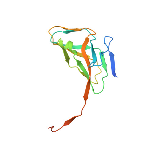
Structures of vaccinia virus dUTPase and its nucleotide complexes.

Samal, A.Schormann, N.Cook, W.J.Delucas, L.J.Chattopadhyay, D.

(2007) Acta Crystallogr D Biol Crystallogr 63: 571-580

  • DOI: https://doi.org/10.1107/S0907444907007871
  • Primary Citation Related Structures: 
    2OKB, 2OKD, 2OKE, 2OL0, 2OL1

  • PubMed Abstract: 

    Deoxyuridine triphosphate nucleotidohydrolase (dUTPase) catalyzes the hydrolysis of dUTP to dUMP and pyrophosphate in the presence of Mg(2+) ions. The enzyme plays multiple cellular roles by maintaining a low dUTP:dTTP ratio and by synthesizing the substrate for thymidylate synthase in the biosynthesis of dTTP. Although dUTPase is an essential enzyme and has been established as a valid target for drug design, the high degree of homology of vaccinia virus dUTPase to the human enzyme makes the identification of selective inhibitors difficult. The crystal structure of vaccinia virus dUTPase has been solved and the active site has been mapped by crystallographic analysis of the apo enzyme and of complexes with the substrate-analog dUMPNPP, with the product dUMP and with dUDP, which acts as an inhibitor. Analyses of these structures reveal subtle differences between the viral and human enzymes. In particular, the much larger size of the central channel at the trimer interface suggests new possibilities for structure-based drug design. Vaccinia virus is a prototype of the poxviruses.


  • Organizational Affiliation
    • Center for Biophysical Sciences and Engineering, University of Alabama at Birmingham, Birmingham, AL 35294, USA.

Macromolecule Content 

  • Total Structure Weight: 49.23 kDa 
  • Atom Count: 3,080 
  • Modeled Residue Count: 372 
  • Deposited Residue Count: 441 
  • Unique protein chains: 1

Macromolecules

Find similar proteins by:|  3D Structure
Entity ID: 1
MoleculeChains  Sequence LengthOrganismDetailsImage
Deoxyuridine 5'-triphosphate nucleotidohydrolase
A, B, C
147Orthopoxvirus vacciniaMutation(s): 1 
Gene Names: DUT
EC: 3.6.1.23
UniProt
Find proteins for P17374 (Vaccinia virus (strain Western Reserve))
Explore P17374 
Go to UniProtKB:  P17374
Entity Groups
Sequence Clusters30% Identity50% Identity70% Identity90% Identity95% Identity100% Identity
UniProt GroupP17374
Sequence Annotations
Expand
Reference Sequence

Experimental Data & Validation

Experimental Data

  • Method: X-RAY DIFFRACTION
  • Resolution: 2.40 Å
  • R-Value Free:  0.268 (Depositor), 0.268 (DCC) 
  • R-Value Work:  0.206 (Depositor), 0.209 (DCC) 
  • R-Value Observed: 0.209 (Depositor) 
Space Group: P 65
Unit Cell:
Length ( Å )Angle ( ˚ )
a = 120.01α = 90
b = 120.01β = 90
c = 50.3γ = 120
Software Package:
Software NamePurpose
REFMACrefinement
DENZOdata reduction
SCALEPACKdata scaling
MOLREPphasing

Structure Validation

View Full Validation Report


Entry History 

Deposition Data

Revision History  (Full details and data files)

  • Version 1.0: 2007-05-01
    Type: Initial release
  • Version 1.1: 2008-05-01
    Changes: Version format compliance
  • Version 1.2: 2011-07-13
    Changes: Advisory, Refinement description, Version format compliance
  • Version 1.3: 2021-10-20
    Changes: Database references, Derived calculations
  • Version 1.4: 2023-08-30
    Changes: Data collection, Refinement description