Discovery of non-covalent dipeptidyl peptidase IV inhibitors which induce a conformational change in the active site.
Sheehan, S.M., Mest, H.J., Watson, B.M., Klimkowski, V.J., Timm, D.E., Cauvin, A., Parsons, S.H., Shi, Q., Canada, E.J., Wiley, M.R., Ruehter, G., Evers, B., Petersen, S., Blaszczak, L.C., Pulley, S.R., Margolis, B.J., Wishart, G.N., Renson, B., Hankotius, D., Mohr, M., Zechel, J.C., Michael Kalbfleisch, J., Dingess-Hammond, E.A., Boelke, A., Weichert, A.G.(2007) Bioorg Med Chem Lett 17: 1765-1768
- PubMed: 17239592 Search on PubMed
- DOI: https://doi.org/10.1016/j.bmcl.2006.12.074
- Primary Citation Related Structures:
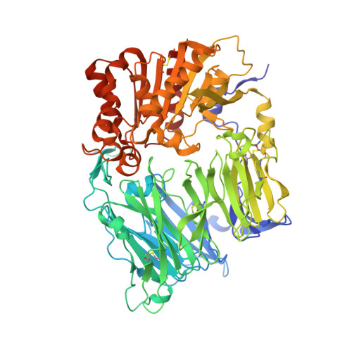
2OGZ - PubMed Abstract:
A series of non-covalent inhibitors of the serine protease dipeptidyl peptidase IV (DPP-IV) were found to adopt a U-shaped binding conformation in X-ray co-crystallization studies. Remarkably, Tyr547 undergoes a 70 degrees side-chain rotation to accommodate the inhibitor and allows access to a previously unexposed area of the protein backbone for hydrogen bonding.
- Lilly Research Laboratories, A Division of Eli Lilly and Company, Indianapolis, IN 46285, USA. SHEEHAN_SCOTT@LILLY.COM
Organizational Affiliation: